Magnesium »
PDB 3bdh-3bre »
3be7 »
Magnesium in PDB 3be7: Crystal Structure of Zn-Dependent Arginine Carboxypeptidase
Protein crystallography data
The structure of Crystal Structure of Zn-Dependent Arginine Carboxypeptidase, PDB code: 3be7
was solved by
Y.Patskovsky,
U.A.Ramagopal,
R.Toro,
A.J.Meyer,
J.Freeman,
M.Iizuka,
K.Bain,
L.Rodgers,
F.Raushel,
J.M.Sauder,
S.K.Burley,
S.C.Almo,
New Yorksgx Research Center For Structural Genomics (Nysgxrc),
with X-Ray Crystallography technique. A brief refinement statistics is given in the table below:
| Resolution Low / High (Å) | 20.00 / 2.30 |
| Space group | P 21 21 21 |
| Cell size a, b, c (Å), α, β, γ (°) | 113.420, 146.583, 255.963, 90.00, 90.00, 90.00 |
| R / Rfree (%) | 22.6 / 28 |
Magnesium Binding Sites:
The binding sites of Magnesium atom in the Crystal Structure of Zn-Dependent Arginine Carboxypeptidase
(pdb code 3be7). This binding sites where shown within
5.0 Angstroms radius around Magnesium atom.
In total 2 binding sites of Magnesium where determined in the Crystal Structure of Zn-Dependent Arginine Carboxypeptidase, PDB code: 3be7:
Jump to Magnesium binding site number: 1; 2;
In total 2 binding sites of Magnesium where determined in the Crystal Structure of Zn-Dependent Arginine Carboxypeptidase, PDB code: 3be7:
Jump to Magnesium binding site number: 1; 2;
Magnesium binding site 1 out of 2 in 3be7
Go back to
Magnesium binding site 1 out
of 2 in the Crystal Structure of Zn-Dependent Arginine Carboxypeptidase
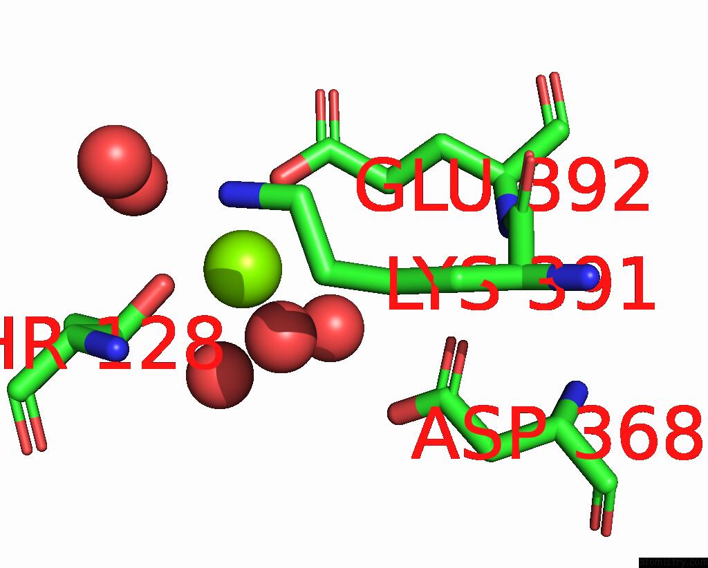
Mono view
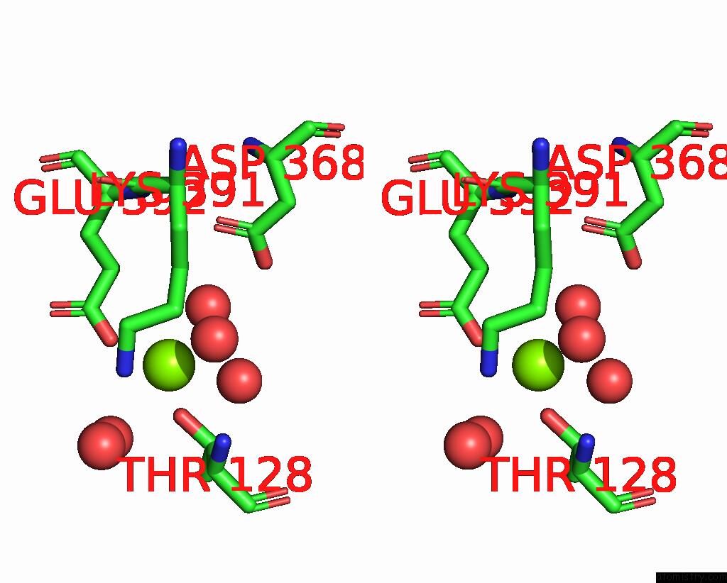
Stereo pair view
Mono view
Stereo pair view
A full contact list of Magnesium with other atoms in the Mg binding
site number 1 of Crystal Structure of Zn-Dependent Arginine Carboxypeptidase within 5.0Å range:
|
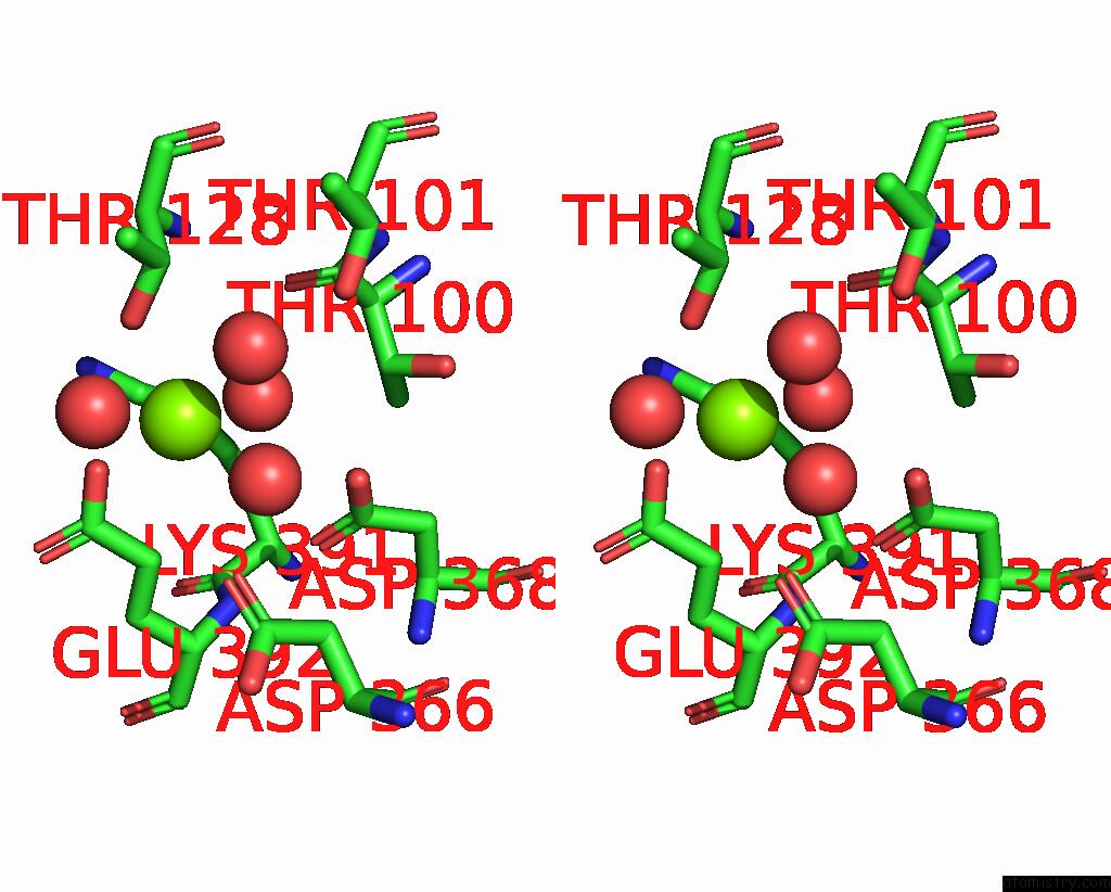
Magnesium binding site 2 out of 2 in 3be7
Go back to
Magnesium binding site 2 out
of 2 in the Crystal Structure of Zn-Dependent Arginine Carboxypeptidase
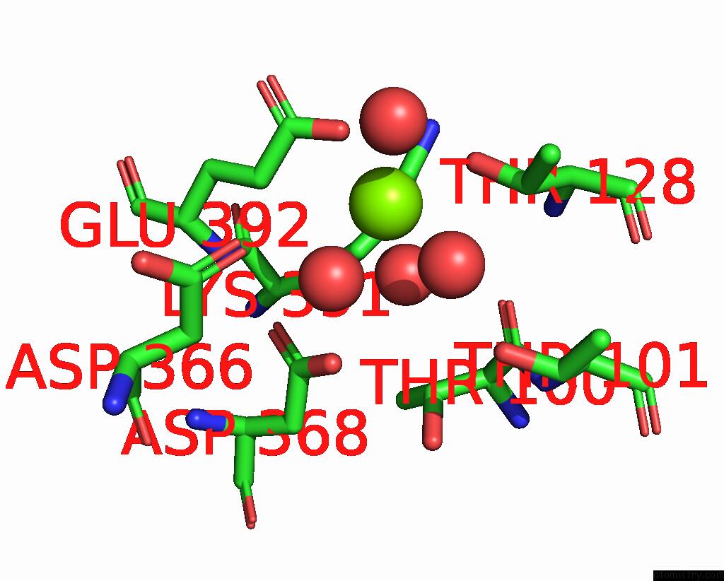
Mono view
Stereo pair view
Mono view
Stereo pair view
A full contact list of Magnesium with other atoms in the Mg binding
site number 2 of Crystal Structure of Zn-Dependent Arginine Carboxypeptidase within 5.0Å range:
|
Reference:
D.F.Xiang,
Y.Patskovsky,
C.Xu,
A.J.Meyer,
J.M.Sauder,
S.K.Burley,
S.C.Almo,
F.M.Raushel.
Functional Identification of Incorrectly Annotated Prolidases From the Amidohydrolase Superfamily of Enzymes. Biochemistry V. 48 3730 2009.
ISSN: ISSN 0006-2960
PubMed: 19281183
DOI: 10.1021/BI900111Q
Page generated: Wed Aug 14 09:09:24 2024
ISSN: ISSN 0006-2960
PubMed: 19281183
DOI: 10.1021/BI900111Q
Last articles
Fe in 2YXOFe in 2YRS
Fe in 2YXC
Fe in 2YNM
Fe in 2YVJ
Fe in 2YP1
Fe in 2YU2
Fe in 2YU1
Fe in 2YQB
Fe in 2YOO