Magnesium »
PDB 3bdh-3bre »
3bjc »
Magnesium in PDB 3bjc: Crystal Structure of the PDE5A Catalytic Domain in Complex with A Novel Inhibitor
Enzymatic activity of Crystal Structure of the PDE5A Catalytic Domain in Complex with A Novel Inhibitor
All present enzymatic activity of Crystal Structure of the PDE5A Catalytic Domain in Complex with A Novel Inhibitor:
3.1.4.35;
3.1.4.35;
Protein crystallography data
The structure of Crystal Structure of the PDE5A Catalytic Domain in Complex with A Novel Inhibitor, PDB code: 3bjc
was solved by
G.Chen,
H.Wang,
R.Howard,
J.Cai,
Y.Wan,
H.Ke,
with X-Ray Crystallography technique. A brief refinement statistics is given in the table below:
| Resolution Low / High (Å) | 30.00 / 2.00 |
| Space group | P 31 2 1 |
| Cell size a, b, c (Å), α, β, γ (°) | 73.755, 73.755, 132.509, 90.00, 90.00, 120.00 |
| R / Rfree (%) | 19.7 / 22.1 |
Other elements in 3bjc:
The structure of Crystal Structure of the PDE5A Catalytic Domain in Complex with A Novel Inhibitor also contains other interesting chemical elements:
| Zinc | (Zn) | 1 atom |
Magnesium Binding Sites:
The binding sites of Magnesium atom in the Crystal Structure of the PDE5A Catalytic Domain in Complex with A Novel Inhibitor
(pdb code 3bjc). This binding sites where shown within
5.0 Angstroms radius around Magnesium atom.
In total only one binding site of Magnesium was determined in the Crystal Structure of the PDE5A Catalytic Domain in Complex with A Novel Inhibitor, PDB code: 3bjc:
In total only one binding site of Magnesium was determined in the Crystal Structure of the PDE5A Catalytic Domain in Complex with A Novel Inhibitor, PDB code: 3bjc:
Magnesium binding site 1 out of 1 in 3bjc
Go back to
Magnesium binding site 1 out
of 1 in the Crystal Structure of the PDE5A Catalytic Domain in Complex with A Novel Inhibitor
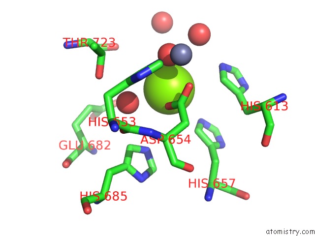
Mono view
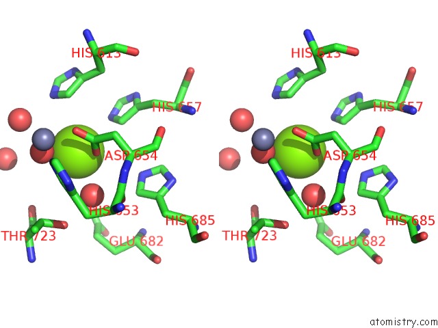
Stereo pair view
Mono view
Stereo pair view
A full contact list of Magnesium with other atoms in the Mg binding
site number 1 of Crystal Structure of the PDE5A Catalytic Domain in Complex with A Novel Inhibitor within 5.0Å range:
|
Reference:
G.Chen,
H.Wang,
H.Robinson,
J.Cai,
Y.Wan,
H.Ke.
An Insight Into the Pharmacophores of Phosphodiesterase-5 Inhibitors From Synthetic and Crystal Structural Studies Biochem.Pharm. V. 75 1717 2008.
ISSN: ISSN 0006-2952
PubMed: 18346713
DOI: 10.1016/J.BCP.2008.01.019
Page generated: Sun Aug 10 17:49:45 2025
ISSN: ISSN 0006-2952
PubMed: 18346713
DOI: 10.1016/J.BCP.2008.01.019
Last articles
Mg in 6QPHMg in 6QV0
Mg in 6QUZ
Mg in 6QUY
Mg in 6QUX
Mg in 6QUW
Mg in 6QUV
Mg in 6QUU
Mg in 6QUS
Mg in 6QTN