Magnesium »
PDB 3bdh-3bre »
3bp1 »
Magnesium in PDB 3bp1: Crystal Structure of Putative 7-Cyano-7-Deazaguanine Reductase Quef From Vibrio Cholerae O1 Biovar Eltor
Enzymatic activity of Crystal Structure of Putative 7-Cyano-7-Deazaguanine Reductase Quef From Vibrio Cholerae O1 Biovar Eltor
All present enzymatic activity of Crystal Structure of Putative 7-Cyano-7-Deazaguanine Reductase Quef From Vibrio Cholerae O1 Biovar Eltor:
1.7.1.13;
1.7.1.13;
Protein crystallography data
The structure of Crystal Structure of Putative 7-Cyano-7-Deazaguanine Reductase Quef From Vibrio Cholerae O1 Biovar Eltor, PDB code: 3bp1
was solved by
Y.Kim,
M.Zhou,
S.Moy,
A.Joachimiak,
Midwest Center For Structuralgenomics (Mcsg),
with X-Ray Crystallography technique. A brief refinement statistics is given in the table below:
| Resolution Low / High (Å) | 26.16 / 1.53 |
| Space group | P 1 |
| Cell size a, b, c (Å), α, β, γ (°) | 71.517, 71.578, 71.511, 119.25, 110.18, 99.58 |
| R / Rfree (%) | 14.3 / 18.3 |
Magnesium Binding Sites:
The binding sites of Magnesium atom in the Crystal Structure of Putative 7-Cyano-7-Deazaguanine Reductase Quef From Vibrio Cholerae O1 Biovar Eltor
(pdb code 3bp1). This binding sites where shown within
5.0 Angstroms radius around Magnesium atom.
In total 2 binding sites of Magnesium where determined in the Crystal Structure of Putative 7-Cyano-7-Deazaguanine Reductase Quef From Vibrio Cholerae O1 Biovar Eltor, PDB code: 3bp1:
Jump to Magnesium binding site number: 1; 2;
In total 2 binding sites of Magnesium where determined in the Crystal Structure of Putative 7-Cyano-7-Deazaguanine Reductase Quef From Vibrio Cholerae O1 Biovar Eltor, PDB code: 3bp1:
Jump to Magnesium binding site number: 1; 2;
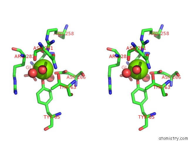
Magnesium binding site 1 out of 2 in 3bp1
Go back to
Magnesium binding site 1 out
of 2 in the Crystal Structure of Putative 7-Cyano-7-Deazaguanine Reductase Quef From Vibrio Cholerae O1 Biovar Eltor
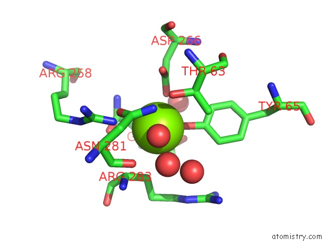
Mono view

Stereo pair view
Mono view
Stereo pair view
A full contact list of Magnesium with other atoms in the Mg binding
site number 1 of Crystal Structure of Putative 7-Cyano-7-Deazaguanine Reductase Quef From Vibrio Cholerae O1 Biovar Eltor within 5.0Å range:
|
Magnesium binding site 2 out of 2 in 3bp1
Go back to
Magnesium binding site 2 out
of 2 in the Crystal Structure of Putative 7-Cyano-7-Deazaguanine Reductase Quef From Vibrio Cholerae O1 Biovar Eltor

Mono view
Stereo pair view
Mono view
Stereo pair view
A full contact list of Magnesium with other atoms in the Mg binding
site number 2 of Crystal Structure of Putative 7-Cyano-7-Deazaguanine Reductase Quef From Vibrio Cholerae O1 Biovar Eltor within 5.0Å range:
|
Reference:
Y.Kim,
M.Zhou,
S.Moy,
J.Morales,
M.A.Cunningham,
A.Joachimiak.
High-Resolution Structure of the Nitrile Reductase Quef Combined with Molecular Simulations Provide Insight Into Enzyme Mechanism. J.Mol.Biol. V. 404 127 2010.
ISSN: ISSN 0022-2836
PubMed: 20875425
DOI: 10.1016/J.JMB.2010.09.042
Page generated: Sun Aug 10 17:54:20 2025
ISSN: ISSN 0022-2836
PubMed: 20875425
DOI: 10.1016/J.JMB.2010.09.042
Last articles
Mg in 6KF9Mg in 6KE2
Mg in 6KF4
Mg in 6KF3
Mg in 6KE4
Mg in 6KE0
Mg in 6KDZ
Mg in 6KDX
Mg in 6KDN
Mg in 6KDM