Magnesium »
PDB 3bdh-3bre »
3bre »
Magnesium in PDB 3bre: Crystal Structure of P.Aeruginosa PA3702
Protein crystallography data
The structure of Crystal Structure of P.Aeruginosa PA3702, PDB code: 3bre
was solved by
N.De,
M.Pirruccello,
P.V.Krasteva,
N.Bae,
R.V.Raghavan,
H.Sondermann,
with X-Ray Crystallography technique. A brief refinement statistics is given in the table below:
| Resolution Low / High (Å) | 33.78 / 2.40 |
| Space group | C 1 2 1 |
| Cell size a, b, c (Å), α, β, γ (°) | 144.510, 72.748, 106.095, 90.00, 110.79, 90.00 |
| R / Rfree (%) | 24.2 / 27.8 |
Magnesium Binding Sites:
The binding sites of Magnesium atom in the Crystal Structure of P.Aeruginosa PA3702
(pdb code 3bre). This binding sites where shown within
5.0 Angstroms radius around Magnesium atom.
In total 2 binding sites of Magnesium where determined in the Crystal Structure of P.Aeruginosa PA3702, PDB code: 3bre:
Jump to Magnesium binding site number: 1; 2;
In total 2 binding sites of Magnesium where determined in the Crystal Structure of P.Aeruginosa PA3702, PDB code: 3bre:
Jump to Magnesium binding site number: 1; 2;
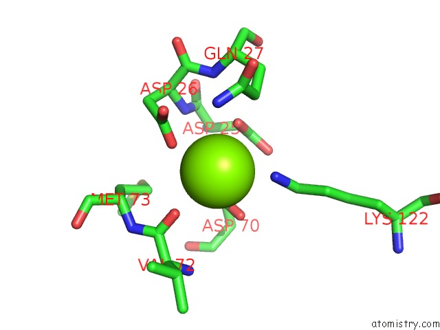
Magnesium binding site 1 out of 2 in 3bre
Go back to
Magnesium binding site 1 out
of 2 in the Crystal Structure of P.Aeruginosa PA3702
Mono view

Stereo pair view
Mono view
Stereo pair view
A full contact list of Magnesium with other atoms in the Mg binding
site number 1 of Crystal Structure of P.Aeruginosa PA3702 within 5.0Å range:
|
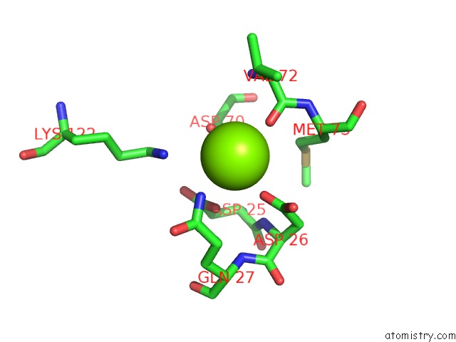
Magnesium binding site 2 out of 2 in 3bre
Go back to
Magnesium binding site 2 out
of 2 in the Crystal Structure of P.Aeruginosa PA3702
Mono view

Stereo pair view
Mono view
Stereo pair view
A full contact list of Magnesium with other atoms in the Mg binding
site number 2 of Crystal Structure of P.Aeruginosa PA3702 within 5.0Å range:
|
Reference:
N.De,
M.Pirruccello,
P.V.Krasteva,
N.Bae,
R.V.Raghavan,
H.Sondermann.
Phosphorylation-Independent Regulation of the Diguanylate Cyclase Wspr. Plos Biol. V. 6 E67 2008.
ISSN: ISSN 1544-9173
PubMed: 18366254
DOI: 10.1371/JOURNAL.PBIO.0060067
Page generated: Sun Aug 10 17:55:26 2025
ISSN: ISSN 1544-9173
PubMed: 18366254
DOI: 10.1371/JOURNAL.PBIO.0060067
Last articles
Mg in 6JWTMg in 6JWU
Mg in 6JWR
Mg in 6JWS
Mg in 6JWP
Mg in 6JWQ
Mg in 6JUP
Mg in 6JUW
Mg in 6JUO
Mg in 6JUN