Magnesium »
PDB 3brw-3c5h »
3bs1 »
Magnesium in PDB 3bs1: Structure of the Staphylococcus Aureus Agra Lyttr Domain Bound to Dna Reveals A Beta Fold with A Novel Mode of Binding
Protein crystallography data
The structure of Structure of the Staphylococcus Aureus Agra Lyttr Domain Bound to Dna Reveals A Beta Fold with A Novel Mode of Binding, PDB code: 3bs1
was solved by
D.J.Sidote,
C.Barbieri,
T.Wu,
A.M.Stock,
with X-Ray Crystallography technique. A brief refinement statistics is given in the table below:
| Resolution Low / High (Å) | 17.30 / 1.60 |
| Space group | P 41 |
| Cell size a, b, c (Å), α, β, γ (°) | 47.938, 47.938, 100.112, 90.00, 90.00, 90.00 |
| R / Rfree (%) | 19.5 / 22.1 |
Other elements in 3bs1:
The structure of Structure of the Staphylococcus Aureus Agra Lyttr Domain Bound to Dna Reveals A Beta Fold with A Novel Mode of Binding also contains other interesting chemical elements:
| Bromine | (Br) | 2 atoms |
Magnesium Binding Sites:
The binding sites of Magnesium atom in the Structure of the Staphylococcus Aureus Agra Lyttr Domain Bound to Dna Reveals A Beta Fold with A Novel Mode of Binding
(pdb code 3bs1). This binding sites where shown within
5.0 Angstroms radius around Magnesium atom.
In total 2 binding sites of Magnesium where determined in the Structure of the Staphylococcus Aureus Agra Lyttr Domain Bound to Dna Reveals A Beta Fold with A Novel Mode of Binding, PDB code: 3bs1:
Jump to Magnesium binding site number: 1; 2;
In total 2 binding sites of Magnesium where determined in the Structure of the Staphylococcus Aureus Agra Lyttr Domain Bound to Dna Reveals A Beta Fold with A Novel Mode of Binding, PDB code: 3bs1:
Jump to Magnesium binding site number: 1; 2;
Magnesium binding site 1 out of 2 in 3bs1
Go back to
Magnesium binding site 1 out
of 2 in the Structure of the Staphylococcus Aureus Agra Lyttr Domain Bound to Dna Reveals A Beta Fold with A Novel Mode of Binding

Mono view
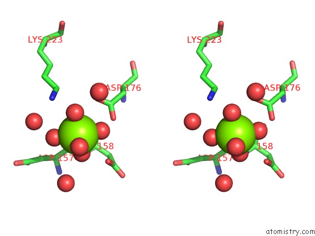
Stereo pair view
Mono view
Stereo pair view
A full contact list of Magnesium with other atoms in the Mg binding
site number 1 of Structure of the Staphylococcus Aureus Agra Lyttr Domain Bound to Dna Reveals A Beta Fold with A Novel Mode of Binding within 5.0Å range:
|
Magnesium binding site 2 out of 2 in 3bs1
Go back to
Magnesium binding site 2 out
of 2 in the Structure of the Staphylococcus Aureus Agra Lyttr Domain Bound to Dna Reveals A Beta Fold with A Novel Mode of Binding

Mono view

Stereo pair view
Mono view
Stereo pair view
A full contact list of Magnesium with other atoms in the Mg binding
site number 2 of Structure of the Staphylococcus Aureus Agra Lyttr Domain Bound to Dna Reveals A Beta Fold with A Novel Mode of Binding within 5.0Å range:
|
Reference:
D.J.Sidote,
C.M.Barbieri,
T.Wu,
A.M.Stock.
Structure of the Staphylococcus Aureus Agra Lyttr Domain Bound to Dna Reveals A Beta Fold with An Unusual Mode of Binding. Structure V. 16 727 2008.
ISSN: ISSN 0969-2126
PubMed: 18462677
DOI: 10.1016/J.STR.2008.02.011
Page generated: Sun Aug 10 17:56:54 2025
ISSN: ISSN 0969-2126
PubMed: 18462677
DOI: 10.1016/J.STR.2008.02.011
Last articles
Mg in 6CA4Mg in 6C90
Mg in 6CA0
Mg in 6C9Y
Mg in 6C8Z
Mg in 6C8P
Mg in 6C8N
Mg in 6C8O
Mg in 6C8D
Mg in 6C8L