Magnesium »
PDB 3brw-3c5h »
3bwy »
Magnesium in PDB 3bwy: Crystal Structure of Human 108M Catechol O-Methyltransferase Bound with S-Adenosylmethionine and Inhibitor Dinitrocatechol
Enzymatic activity of Crystal Structure of Human 108M Catechol O-Methyltransferase Bound with S-Adenosylmethionine and Inhibitor Dinitrocatechol
All present enzymatic activity of Crystal Structure of Human 108M Catechol O-Methyltransferase Bound with S-Adenosylmethionine and Inhibitor Dinitrocatechol:
2.1.1.6;
2.1.1.6;
Protein crystallography data
The structure of Crystal Structure of Human 108M Catechol O-Methyltransferase Bound with S-Adenosylmethionine and Inhibitor Dinitrocatechol, PDB code: 3bwy
was solved by
K.Rutherford,
I.Le Trong,
R.E.Stenkamp,
W.W.Parson,
with X-Ray Crystallography technique. A brief refinement statistics is given in the table below:
| Resolution Low / High (Å) | 47.62 / 1.30 |
| Space group | P 21 21 21 |
| Cell size a, b, c (Å), α, β, γ (°) | 43.249, 66.163, 68.519, 90.00, 90.00, 90.00 |
| R / Rfree (%) | 12.4 / 16.7 |
Magnesium Binding Sites:
The binding sites of Magnesium atom in the Crystal Structure of Human 108M Catechol O-Methyltransferase Bound with S-Adenosylmethionine and Inhibitor Dinitrocatechol
(pdb code 3bwy). This binding sites where shown within
5.0 Angstroms radius around Magnesium atom.
In total only one binding site of Magnesium was determined in the Crystal Structure of Human 108M Catechol O-Methyltransferase Bound with S-Adenosylmethionine and Inhibitor Dinitrocatechol, PDB code: 3bwy:
In total only one binding site of Magnesium was determined in the Crystal Structure of Human 108M Catechol O-Methyltransferase Bound with S-Adenosylmethionine and Inhibitor Dinitrocatechol, PDB code: 3bwy:
Magnesium binding site 1 out of 1 in 3bwy
Go back to
Magnesium binding site 1 out
of 1 in the Crystal Structure of Human 108M Catechol O-Methyltransferase Bound with S-Adenosylmethionine and Inhibitor Dinitrocatechol
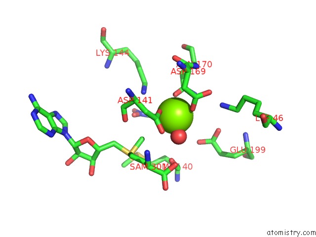
Mono view
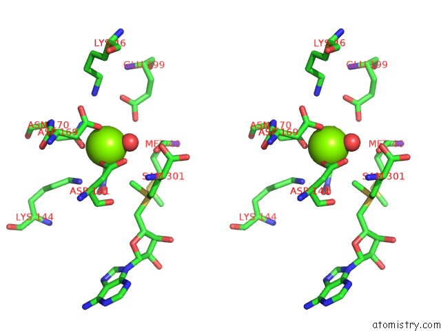
Stereo pair view
Mono view
Stereo pair view
A full contact list of Magnesium with other atoms in the Mg binding
site number 1 of Crystal Structure of Human 108M Catechol O-Methyltransferase Bound with S-Adenosylmethionine and Inhibitor Dinitrocatechol within 5.0Å range:
|
Reference:
K.Rutherford,
I.Le Trong,
R.E.Stenkamp,
W.W.Parson.
Crystal Structures of Human 108V and 108M Catechol O-Methyltransferase. J.Mol.Biol. V. 380 120 2008.
ISSN: ISSN 0022-2836
PubMed: 18486144
DOI: 10.1016/J.JMB.2008.04.040
Page generated: Sun Aug 10 17:58:20 2025
ISSN: ISSN 0022-2836
PubMed: 18486144
DOI: 10.1016/J.JMB.2008.04.040
Last articles
Mg in 6CA4Mg in 6C90
Mg in 6CA0
Mg in 6C9Y
Mg in 6C8Z
Mg in 6C8P
Mg in 6C8N
Mg in 6C8O
Mg in 6C8D
Mg in 6C8L