Magnesium »
PDB 3brw-3c5h »
3bxz »
Magnesium in PDB 3bxz: Crystal Structure of the Isolated Dead Motor Domains From Escherichia Coli Seca
Protein crystallography data
The structure of Crystal Structure of the Isolated Dead Motor Domains From Escherichia Coli Seca, PDB code: 3bxz
was solved by
S.Nithianantham,
S.Namjoshi,
B.H.Shilton,
with X-Ray Crystallography technique. A brief refinement statistics is given in the table below:
| Resolution Low / High (Å) | 46.58 / 3.00 |
| Space group | C 1 2 1 |
| Cell size a, b, c (Å), α, β, γ (°) | 177.823, 71.361, 119.536, 90.00, 128.82, 90.00 |
| R / Rfree (%) | 24.5 / 30.5 |
Magnesium Binding Sites:
The binding sites of Magnesium atom in the Crystal Structure of the Isolated Dead Motor Domains From Escherichia Coli Seca
(pdb code 3bxz). This binding sites where shown within
5.0 Angstroms radius around Magnesium atom.
In total 2 binding sites of Magnesium where determined in the Crystal Structure of the Isolated Dead Motor Domains From Escherichia Coli Seca, PDB code: 3bxz:
Jump to Magnesium binding site number: 1; 2;
In total 2 binding sites of Magnesium where determined in the Crystal Structure of the Isolated Dead Motor Domains From Escherichia Coli Seca, PDB code: 3bxz:
Jump to Magnesium binding site number: 1; 2;
Magnesium binding site 1 out of 2 in 3bxz
Go back to
Magnesium binding site 1 out
of 2 in the Crystal Structure of the Isolated Dead Motor Domains From Escherichia Coli Seca
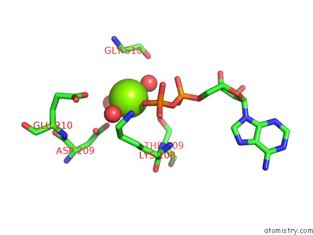
Mono view
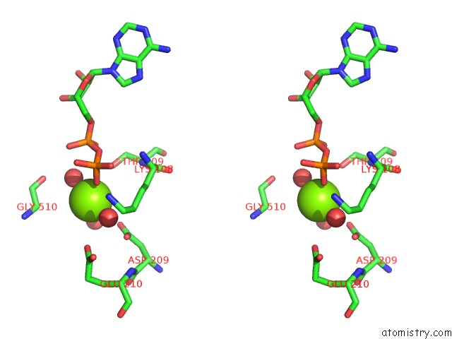
Stereo pair view
Mono view
Stereo pair view
A full contact list of Magnesium with other atoms in the Mg binding
site number 1 of Crystal Structure of the Isolated Dead Motor Domains From Escherichia Coli Seca within 5.0Å range:
|
Magnesium binding site 2 out of 2 in 3bxz
Go back to
Magnesium binding site 2 out
of 2 in the Crystal Structure of the Isolated Dead Motor Domains From Escherichia Coli Seca
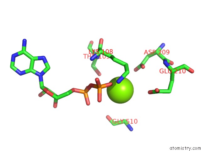
Mono view
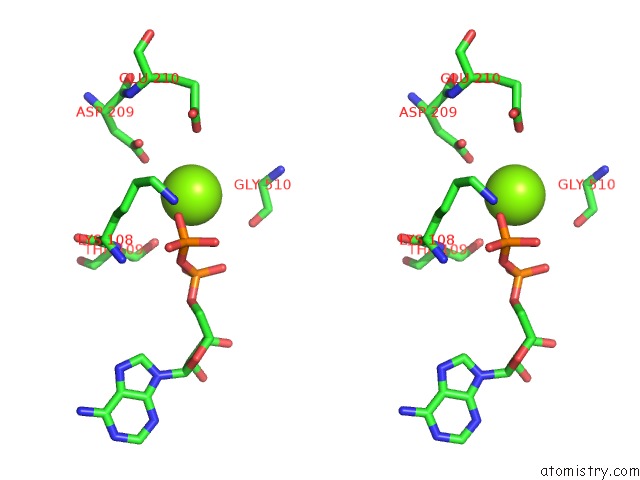
Stereo pair view
Mono view
Stereo pair view
A full contact list of Magnesium with other atoms in the Mg binding
site number 2 of Crystal Structure of the Isolated Dead Motor Domains From Escherichia Coli Seca within 5.0Å range:
|
Reference:
S.Nithianantham,
B.H.Shilton.
Analysis of the Isolated Seca Dead Motor Suggests A Mechanism For Chemical-Mechanical Coupling. J.Mol.Biol. V. 383 380 2008.
ISSN: ISSN 0022-2836
PubMed: 18761349
DOI: 10.1016/J.JMB.2008.08.022
Page generated: Sun Aug 10 17:58:28 2025
ISSN: ISSN 0022-2836
PubMed: 18761349
DOI: 10.1016/J.JMB.2008.08.022
Last articles
Mg in 6CA4Mg in 6C90
Mg in 6CA0
Mg in 6C9Y
Mg in 6C8Z
Mg in 6C8P
Mg in 6C8N
Mg in 6C8O
Mg in 6C8D
Mg in 6C8L