Magnesium »
PDB 3brw-3c5h »
3c3c »
Magnesium in PDB 3c3c: Crystal Structure of Human Phosphoglycerate Kinase Bound to 3-Phosphoglycerate and L-Cdp
Enzymatic activity of Crystal Structure of Human Phosphoglycerate Kinase Bound to 3-Phosphoglycerate and L-Cdp
All present enzymatic activity of Crystal Structure of Human Phosphoglycerate Kinase Bound to 3-Phosphoglycerate and L-Cdp:
2.7.2.3;
2.7.2.3;
Protein crystallography data
The structure of Crystal Structure of Human Phosphoglycerate Kinase Bound to 3-Phosphoglycerate and L-Cdp, PDB code: 3c3c
was solved by
S.T.Arold,
C.Gondeau,
C.Lionne,
L.Chaloin,
with X-Ray Crystallography technique. A brief refinement statistics is given in the table below:
| Resolution Low / High (Å) | 43.60 / 2.40 |
| Space group | P 1 |
| Cell size a, b, c (Å), α, β, γ (°) | 35.789, 55.824, 94.497, 78.95, 84.51, 83.23 |
| R / Rfree (%) | 19.3 / 28.1 |
Magnesium Binding Sites:
The binding sites of Magnesium atom in the Crystal Structure of Human Phosphoglycerate Kinase Bound to 3-Phosphoglycerate and L-Cdp
(pdb code 3c3c). This binding sites where shown within
5.0 Angstroms radius around Magnesium atom.
In total only one binding site of Magnesium was determined in the Crystal Structure of Human Phosphoglycerate Kinase Bound to 3-Phosphoglycerate and L-Cdp, PDB code: 3c3c:
In total only one binding site of Magnesium was determined in the Crystal Structure of Human Phosphoglycerate Kinase Bound to 3-Phosphoglycerate and L-Cdp, PDB code: 3c3c:
Magnesium binding site 1 out of 1 in 3c3c
Go back to
Magnesium binding site 1 out
of 1 in the Crystal Structure of Human Phosphoglycerate Kinase Bound to 3-Phosphoglycerate and L-Cdp
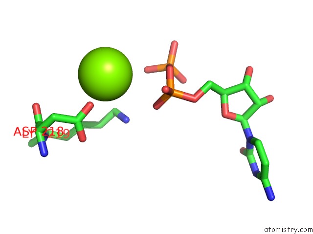
Mono view
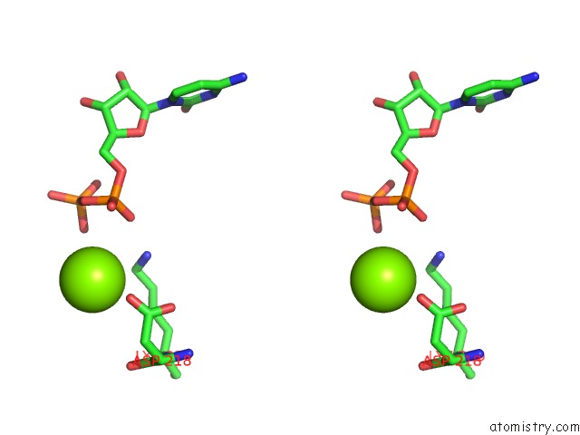
Stereo pair view
Mono view
Stereo pair view
A full contact list of Magnesium with other atoms in the Mg binding
site number 1 of Crystal Structure of Human Phosphoglycerate Kinase Bound to 3-Phosphoglycerate and L-Cdp within 5.0Å range:
|
Reference:
C.Gondeau,
L.Chaloin,
P.Lallemand,
B.Roy,
C.Perigaud,
T.Barman,
A.Varga,
M.Vas,
C.Lionne,
S.T.Arold.
Molecular Basis For the Lack of Enantioselectivity of Human 3-Phosphoglycerate Kinase Nucleic Acids Res. V. 36 3620 2008.
ISSN: ISSN 0305-1048
PubMed: 18463139
DOI: 10.1093/NAR/GKN212
Page generated: Sun Aug 10 17:59:43 2025
ISSN: ISSN 0305-1048
PubMed: 18463139
DOI: 10.1093/NAR/GKN212
Last articles
Mg in 7GYDMg in 7FSD
Mg in 7GYC
Mg in 7GYB
Mg in 7GYA
Mg in 7GY8
Mg in 7GY9
Mg in 7GY7
Mg in 7GY6
Mg in 7GY5