Magnesium »
PDB 3brw-3c5h »
3c4v »
Magnesium in PDB 3c4v: Structure of the Retaining Glycosyltransferase Msha:the First Step in Mycothiol Biosynthesis. Organism: Corynebacterium Glutamicum : Complex with Udp and 1L-Ins-1- P.
Protein crystallography data
The structure of Structure of the Retaining Glycosyltransferase Msha:the First Step in Mycothiol Biosynthesis. Organism: Corynebacterium Glutamicum : Complex with Udp and 1L-Ins-1- P., PDB code: 3c4v
was solved by
M.W.Vetting,
P.A.Frantom,
J.S.Blanchard,
with X-Ray Crystallography technique. A brief refinement statistics is given in the table below:
| Resolution Low / High (Å) | 79.18 / 2.60 |
| Space group | I 4 2 2 |
| Cell size a, b, c (Å), α, β, γ (°) | 223.940, 223.940, 125.005, 90.00, 90.00, 90.00 |
| R / Rfree (%) | 19 / 21.3 |
Magnesium Binding Sites:
The binding sites of Magnesium atom in the Structure of the Retaining Glycosyltransferase Msha:the First Step in Mycothiol Biosynthesis. Organism: Corynebacterium Glutamicum : Complex with Udp and 1L-Ins-1- P.
(pdb code 3c4v). This binding sites where shown within
5.0 Angstroms radius around Magnesium atom.
In total 2 binding sites of Magnesium where determined in the Structure of the Retaining Glycosyltransferase Msha:the First Step in Mycothiol Biosynthesis. Organism: Corynebacterium Glutamicum : Complex with Udp and 1L-Ins-1- P., PDB code: 3c4v:
Jump to Magnesium binding site number: 1; 2;
In total 2 binding sites of Magnesium where determined in the Structure of the Retaining Glycosyltransferase Msha:the First Step in Mycothiol Biosynthesis. Organism: Corynebacterium Glutamicum : Complex with Udp and 1L-Ins-1- P., PDB code: 3c4v:
Jump to Magnesium binding site number: 1; 2;
Magnesium binding site 1 out of 2 in 3c4v
Go back to
Magnesium binding site 1 out
of 2 in the Structure of the Retaining Glycosyltransferase Msha:the First Step in Mycothiol Biosynthesis. Organism: Corynebacterium Glutamicum : Complex with Udp and 1L-Ins-1- P.
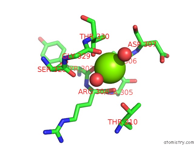
Mono view
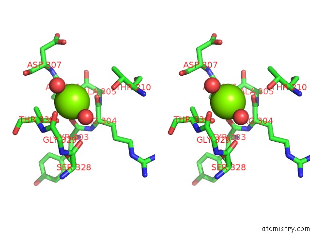
Stereo pair view
Mono view
Stereo pair view
A full contact list of Magnesium with other atoms in the Mg binding
site number 1 of Structure of the Retaining Glycosyltransferase Msha:the First Step in Mycothiol Biosynthesis. Organism: Corynebacterium Glutamicum : Complex with Udp and 1L-Ins-1- P. within 5.0Å range:
|
Magnesium binding site 2 out of 2 in 3c4v
Go back to
Magnesium binding site 2 out
of 2 in the Structure of the Retaining Glycosyltransferase Msha:the First Step in Mycothiol Biosynthesis. Organism: Corynebacterium Glutamicum : Complex with Udp and 1L-Ins-1- P.
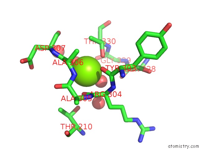
Mono view
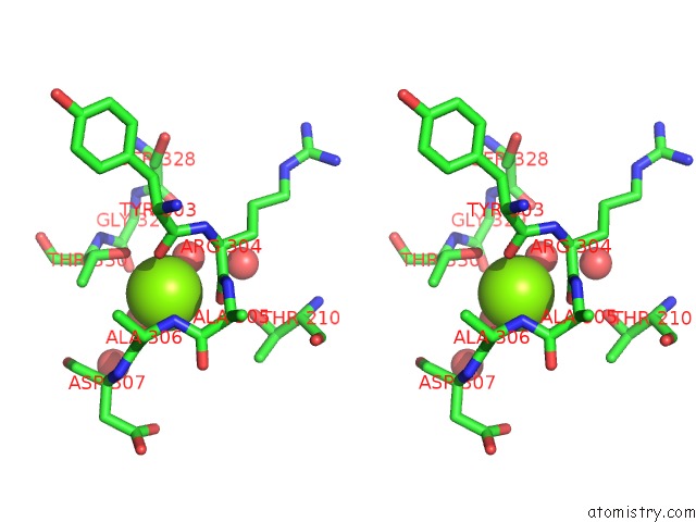
Stereo pair view
Mono view
Stereo pair view
A full contact list of Magnesium with other atoms in the Mg binding
site number 2 of Structure of the Retaining Glycosyltransferase Msha:the First Step in Mycothiol Biosynthesis. Organism: Corynebacterium Glutamicum : Complex with Udp and 1L-Ins-1- P. within 5.0Å range:
|
Reference:
M.W.Vetting,
P.A.Frantom,
J.S.Blanchard.
Structural and Enzymatic Analysis of Msha From Corynebacterium Glutamicum: Substrate-Assisted Catalysis J.Biol.Chem. V. 283 15834 2008.
ISSN: ISSN 0021-9258
PubMed: 18390549
DOI: 10.1074/JBC.M801017200
Page generated: Sun Aug 10 18:00:19 2025
ISSN: ISSN 0021-9258
PubMed: 18390549
DOI: 10.1074/JBC.M801017200
Last articles
Mg in 6CA4Mg in 6C90
Mg in 6CA0
Mg in 6C9Y
Mg in 6C8Z
Mg in 6C8P
Mg in 6C8N
Mg in 6C8O
Mg in 6C8D
Mg in 6C8L