Magnesium »
PDB 3c5i-3cf5 »
3c6a »
Magnesium in PDB 3c6a: Crystal Structure of the RB49 GP17 Nuclease Domain
Protein crystallography data
The structure of Crystal Structure of the RB49 GP17 Nuclease Domain, PDB code: 3c6a
was solved by
S.Sun,
M.G.Rossmann,
with X-Ray Crystallography technique. A brief refinement statistics is given in the table below:
| Resolution Low / High (Å) | 10.00 / 1.16 |
| Space group | P 21 21 2 |
| Cell size a, b, c (Å), α, β, γ (°) | 52.568, 125.163, 37.218, 90.00, 90.00, 90.00 |
| R / Rfree (%) | 13.4 / 17.1 |
Magnesium Binding Sites:
The binding sites of Magnesium atom in the Crystal Structure of the RB49 GP17 Nuclease Domain
(pdb code 3c6a). This binding sites where shown within
5.0 Angstroms radius around Magnesium atom.
In total only one binding site of Magnesium was determined in the Crystal Structure of the RB49 GP17 Nuclease Domain, PDB code: 3c6a:
In total only one binding site of Magnesium was determined in the Crystal Structure of the RB49 GP17 Nuclease Domain, PDB code: 3c6a:
Magnesium binding site 1 out of 1 in 3c6a
Go back to
Magnesium binding site 1 out
of 1 in the Crystal Structure of the RB49 GP17 Nuclease Domain
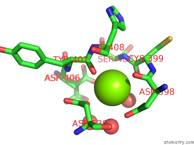
Mono view
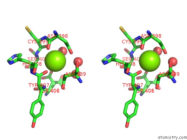
Stereo pair view
Mono view
Stereo pair view
A full contact list of Magnesium with other atoms in the Mg binding
site number 1 of Crystal Structure of the RB49 GP17 Nuclease Domain within 5.0Å range:
|
Reference:
S.Sun,
K.Kondabagil,
B.Draper,
T.I.Alam,
V.D.Bowman,
Z.Zhang,
S.Hegde,
A.Fokine,
M.G.Rossmann,
V.B.Rao.
The Structure of the Phage T4 Dna Packaging Motor Suggests A Mechanism Dependent on Electrostatic Forces. Cell(Cambridge,Mass.) V. 135 1251 2008.
ISSN: ISSN 0092-8674
PubMed: 19109896
DOI: 10.1016/J.CELL.2008.11.015
Page generated: Wed Aug 14 09:29:13 2024
ISSN: ISSN 0092-8674
PubMed: 19109896
DOI: 10.1016/J.CELL.2008.11.015
Last articles
Zn in 9J0NZn in 9J0O
Zn in 9J0P
Zn in 9FJX
Zn in 9EKB
Zn in 9C0F
Zn in 9CAH
Zn in 9CH0
Zn in 9CH3
Zn in 9CH1