Magnesium »
PDB 3c5i-3cf5 »
3caw »
Magnesium in PDB 3caw: Crystal Structure of O-Succinylbenzoate Synthase From Bdellovibrio Bacteriovorus Liganded with Mg
Protein crystallography data
The structure of Crystal Structure of O-Succinylbenzoate Synthase From Bdellovibrio Bacteriovorus Liganded with Mg, PDB code: 3caw
was solved by
A.A.Fedorov,
E.V.Fedorov,
A.Sakai,
S.K.Burley,
J.A.Gerlt,
S.C.Almo,
Newyork Sgx Research Center For Structural Genomics (Nysgxrc),
with X-Ray Crystallography technique. A brief refinement statistics is given in the table below:
| Resolution Low / High (Å) | 24.85 / 1.87 |
| Space group | P 1 21 1 |
| Cell size a, b, c (Å), α, β, γ (°) | 53.381, 78.610, 78.462, 90.00, 91.78, 90.00 |
| R / Rfree (%) | 21.8 / 24.4 |
Magnesium Binding Sites:
The binding sites of Magnesium atom in the Crystal Structure of O-Succinylbenzoate Synthase From Bdellovibrio Bacteriovorus Liganded with Mg
(pdb code 3caw). This binding sites where shown within
5.0 Angstroms radius around Magnesium atom.
In total 2 binding sites of Magnesium where determined in the Crystal Structure of O-Succinylbenzoate Synthase From Bdellovibrio Bacteriovorus Liganded with Mg, PDB code: 3caw:
Jump to Magnesium binding site number: 1; 2;
In total 2 binding sites of Magnesium where determined in the Crystal Structure of O-Succinylbenzoate Synthase From Bdellovibrio Bacteriovorus Liganded with Mg, PDB code: 3caw:
Jump to Magnesium binding site number: 1; 2;
Magnesium binding site 1 out of 2 in 3caw
Go back to
Magnesium binding site 1 out
of 2 in the Crystal Structure of O-Succinylbenzoate Synthase From Bdellovibrio Bacteriovorus Liganded with Mg
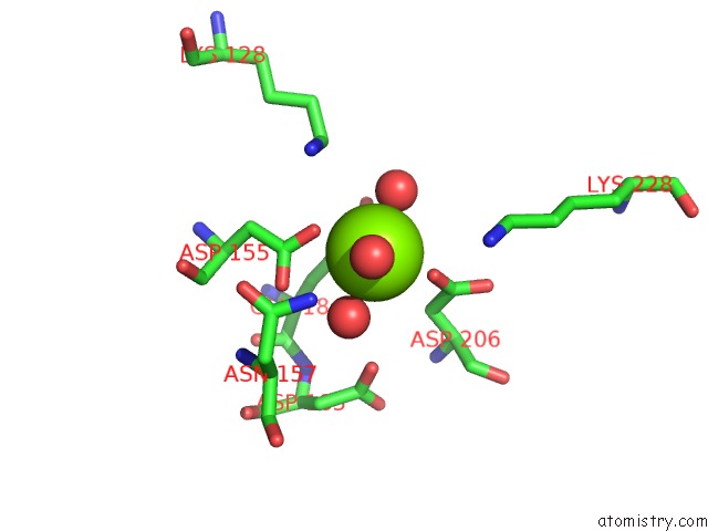
Mono view
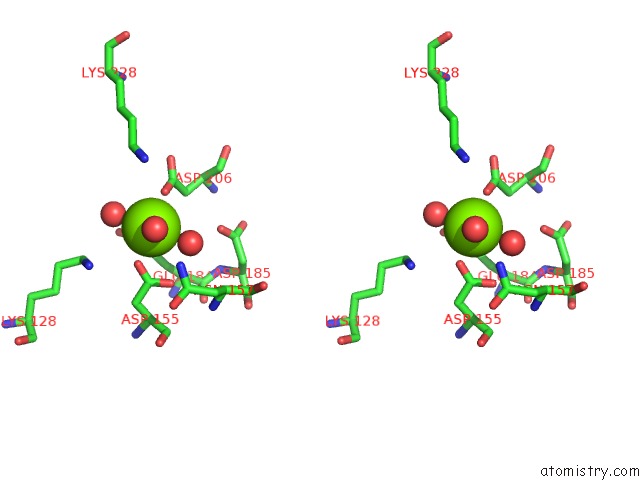
Stereo pair view
Mono view
Stereo pair view
A full contact list of Magnesium with other atoms in the Mg binding
site number 1 of Crystal Structure of O-Succinylbenzoate Synthase From Bdellovibrio Bacteriovorus Liganded with Mg within 5.0Å range:
|
Magnesium binding site 2 out of 2 in 3caw
Go back to
Magnesium binding site 2 out
of 2 in the Crystal Structure of O-Succinylbenzoate Synthase From Bdellovibrio Bacteriovorus Liganded with Mg
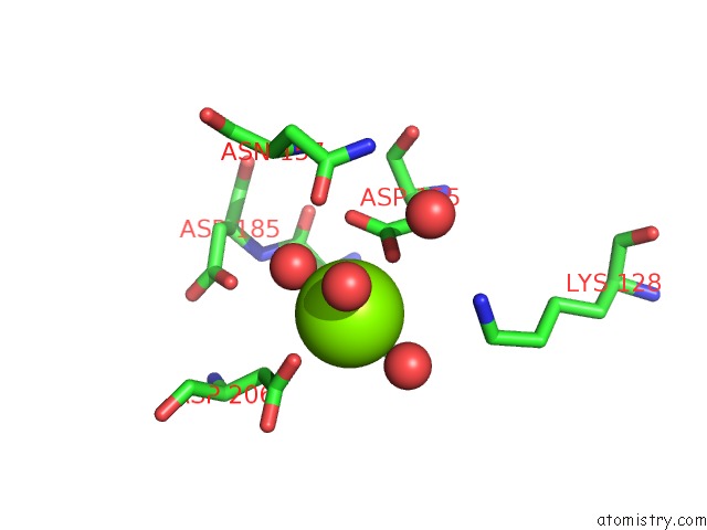
Mono view
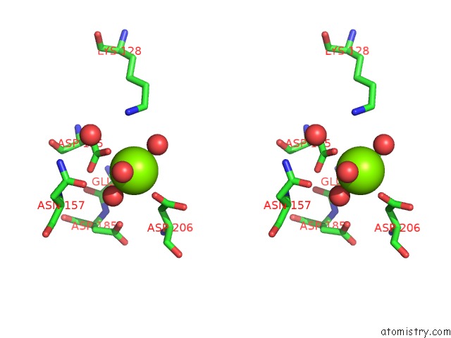
Stereo pair view
Mono view
Stereo pair view
A full contact list of Magnesium with other atoms in the Mg binding
site number 2 of Crystal Structure of O-Succinylbenzoate Synthase From Bdellovibrio Bacteriovorus Liganded with Mg within 5.0Å range:
|
Reference:
D.Odokonyero,
A.Sakai,
Y.Patskovsky,
V.N.Malashkevich,
A.A.Fedorov,
J.B.Bonanno,
E.V.Fedorov,
R.Toro,
R.Agarwal,
C.Wang,
N.D.Ozerova,
W.S.Yew,
J.M.Sauder,
S.Swaminathan,
S.K.Burley,
S.C.Almo,
M.E.Glasner.
Loss of Quaternary Structure Is Associated with Rapid Sequence Divergence in the Osbs Family. Proc.Natl.Acad.Sci.Usa V. 111 8535 2014.
ISSN: ISSN 0027-8424
PubMed: 24872444
DOI: 10.1073/PNAS.1318703111
Page generated: Wed Aug 14 09:32:23 2024
ISSN: ISSN 0027-8424
PubMed: 24872444
DOI: 10.1073/PNAS.1318703111
Last articles
Fe in 2YXOFe in 2YRS
Fe in 2YXC
Fe in 2YNM
Fe in 2YVJ
Fe in 2YP1
Fe in 2YU2
Fe in 2YU1
Fe in 2YQB
Fe in 2YOO