Magnesium »
PDB 3cfx-3cpj »
3cfx »
Magnesium in PDB 3cfx: Crystal Structure of M. Acetivorans Periplasmic Binding Protein Moda/Wtpa with Bound Tungstate
Protein crystallography data
The structure of Crystal Structure of M. Acetivorans Periplasmic Binding Protein Moda/Wtpa with Bound Tungstate, PDB code: 3cfx
was solved by
M.Comellas-Bigler,
K.Hollenstein,
K.P.Locher,
with X-Ray Crystallography technique. A brief refinement statistics is given in the table below:
| Resolution Low / High (Å) | 30.00 / 1.60 |
| Space group | C 1 2 1 |
| Cell size a, b, c (Å), α, β, γ (°) | 115.500, 51.860, 124.048, 90.00, 116.93, 90.00 |
| R / Rfree (%) | 21 / 23.5 |
Other elements in 3cfx:
The structure of Crystal Structure of M. Acetivorans Periplasmic Binding Protein Moda/Wtpa with Bound Tungstate also contains other interesting chemical elements:
| Tungsten | (W) | 2 atoms |
Magnesium Binding Sites:
The binding sites of Magnesium atom in the Crystal Structure of M. Acetivorans Periplasmic Binding Protein Moda/Wtpa with Bound Tungstate
(pdb code 3cfx). This binding sites where shown within
5.0 Angstroms radius around Magnesium atom.
In total 4 binding sites of Magnesium where determined in the Crystal Structure of M. Acetivorans Periplasmic Binding Protein Moda/Wtpa with Bound Tungstate, PDB code: 3cfx:
Jump to Magnesium binding site number: 1; 2; 3; 4;
In total 4 binding sites of Magnesium where determined in the Crystal Structure of M. Acetivorans Periplasmic Binding Protein Moda/Wtpa with Bound Tungstate, PDB code: 3cfx:
Jump to Magnesium binding site number: 1; 2; 3; 4;
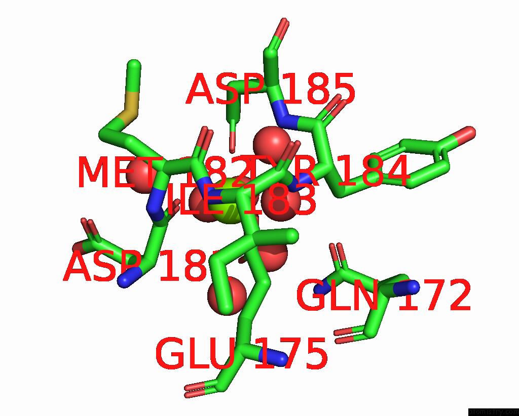
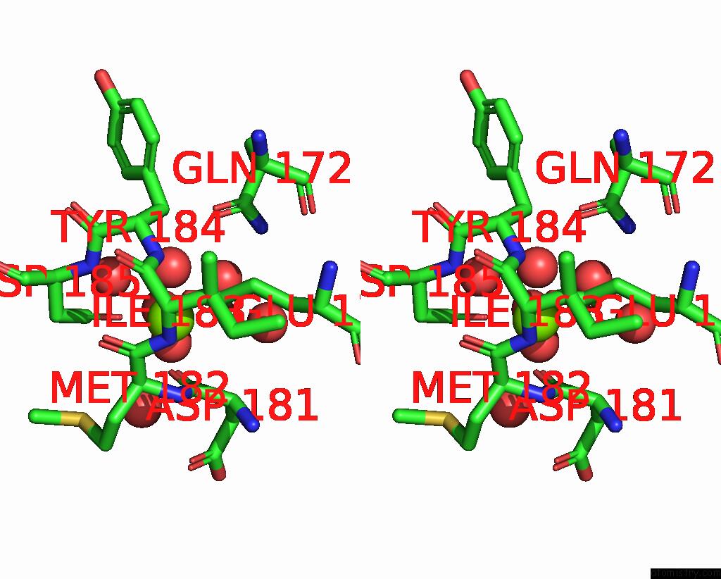
Magnesium binding site 1 out of 4 in 3cfx
Go back to
Magnesium binding site 1 out
of 4 in the Crystal Structure of M. Acetivorans Periplasmic Binding Protein Moda/Wtpa with Bound Tungstate
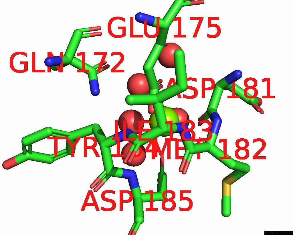
Mono view
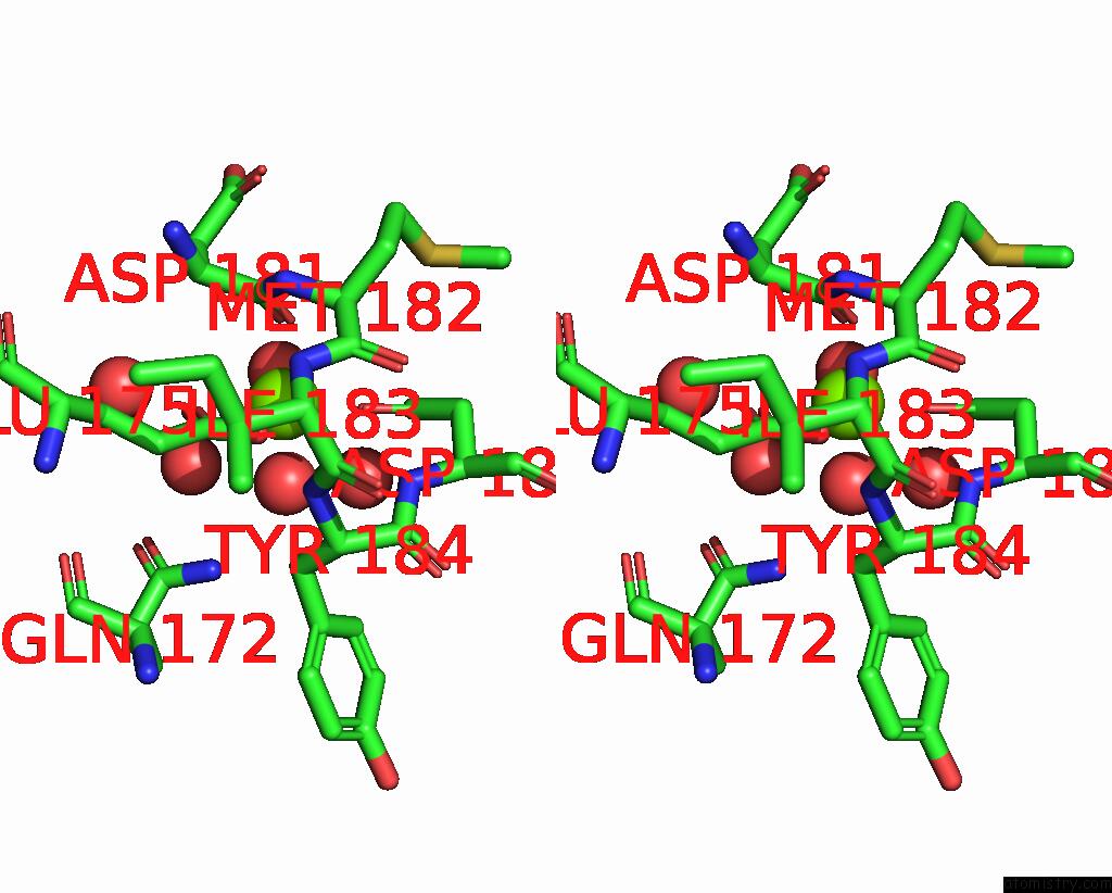
Stereo pair view
Mono view
Stereo pair view
A full contact list of Magnesium with other atoms in the Mg binding
site number 1 of Crystal Structure of M. Acetivorans Periplasmic Binding Protein Moda/Wtpa with Bound Tungstate within 5.0Å range:
|
Magnesium binding site 2 out of 4 in 3cfx
Go back to
Magnesium binding site 2 out
of 4 in the Crystal Structure of M. Acetivorans Periplasmic Binding Protein Moda/Wtpa with Bound Tungstate
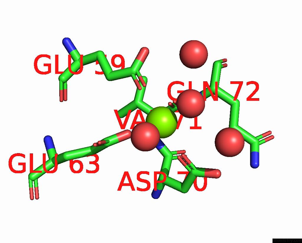
Mono view
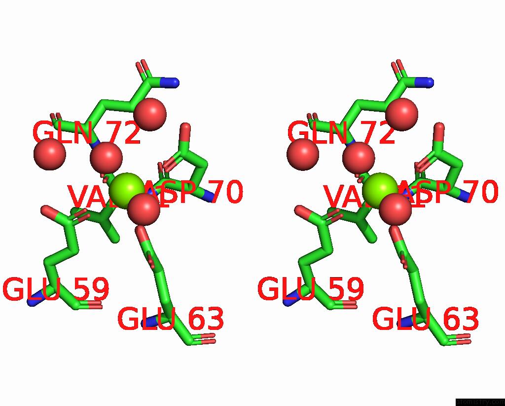
Stereo pair view
Mono view
Stereo pair view
A full contact list of Magnesium with other atoms in the Mg binding
site number 2 of Crystal Structure of M. Acetivorans Periplasmic Binding Protein Moda/Wtpa with Bound Tungstate within 5.0Å range:
|
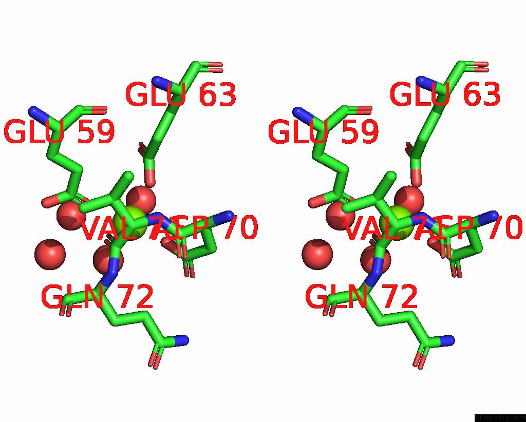
Magnesium binding site 3 out of 4 in 3cfx
Go back to
Magnesium binding site 3 out
of 4 in the Crystal Structure of M. Acetivorans Periplasmic Binding Protein Moda/Wtpa with Bound Tungstate
Mono view
Stereo pair view
Mono view
Stereo pair view
A full contact list of Magnesium with other atoms in the Mg binding
site number 3 of Crystal Structure of M. Acetivorans Periplasmic Binding Protein Moda/Wtpa with Bound Tungstate within 5.0Å range:
|
Magnesium binding site 4 out of 4 in 3cfx
Go back to
Magnesium binding site 4 out
of 4 in the Crystal Structure of M. Acetivorans Periplasmic Binding Protein Moda/Wtpa with Bound Tungstate

Mono view
Stereo pair view
Mono view
Stereo pair view
A full contact list of Magnesium with other atoms in the Mg binding
site number 4 of Crystal Structure of M. Acetivorans Periplasmic Binding Protein Moda/Wtpa with Bound Tungstate within 5.0Å range:
|
Reference:
K.Hollenstein,
M.Comellas-Bigler,
L.E.Bevers,
M.C.Feiters,
W.Meyer-Klaucke,
P.L.Hagedoorn,
K.P.Locher.
Distorted Octahedral Coordination of Tungstate in A Subfamily of Specific Binding Proteins. J.Biol.Inorg.Chem. V. 14 663 2009.
ISSN: ISSN 0949-8257
PubMed: 19234723
DOI: 10.1007/S00775-009-0479-7
Page generated: Sun Aug 10 19:21:35 2025
ISSN: ISSN 0949-8257
PubMed: 19234723
DOI: 10.1007/S00775-009-0479-7
Last articles
Mg in 3I5XMg in 3I4K
Mg in 3I5F
Mg in 3I5C
Mg in 3I4N
Mg in 3I3E
Mg in 3I4D
Mg in 3I4M
Mg in 3I3D
Mg in 3I3B