Magnesium »
PDB 3cfx-3cpj »
3cgs »
Magnesium in PDB 3cgs: X-Ray Structure Containing the Pseudouridylated U2 Snrna and Mammalian Intron Branch Site Consensus Sequences
Protein crystallography data
The structure of X-Ray Structure Containing the Pseudouridylated U2 Snrna and Mammalian Intron Branch Site Consensus Sequences, PDB code: 3cgs
was solved by
Y.Lin,
C.L.Kielkopf,
with X-Ray Crystallography technique. A brief refinement statistics is given in the table below:
| Resolution Low / High (Å) | 20.00 / 1.65 |
| Space group | P 1 21 1 |
| Cell size a, b, c (Å), α, β, γ (°) | 31.190, 38.280, 32.080, 90.00, 108.23, 90.00 |
| R / Rfree (%) | 22.7 / 25.2 |
Magnesium Binding Sites:
The binding sites of Magnesium atom in the X-Ray Structure Containing the Pseudouridylated U2 Snrna and Mammalian Intron Branch Site Consensus Sequences
(pdb code 3cgs). This binding sites where shown within
5.0 Angstroms radius around Magnesium atom.
In total only one binding site of Magnesium was determined in the X-Ray Structure Containing the Pseudouridylated U2 Snrna and Mammalian Intron Branch Site Consensus Sequences, PDB code: 3cgs:
In total only one binding site of Magnesium was determined in the X-Ray Structure Containing the Pseudouridylated U2 Snrna and Mammalian Intron Branch Site Consensus Sequences, PDB code: 3cgs:
Magnesium binding site 1 out of 1 in 3cgs
Go back to
Magnesium binding site 1 out
of 1 in the X-Ray Structure Containing the Pseudouridylated U2 Snrna and Mammalian Intron Branch Site Consensus Sequences
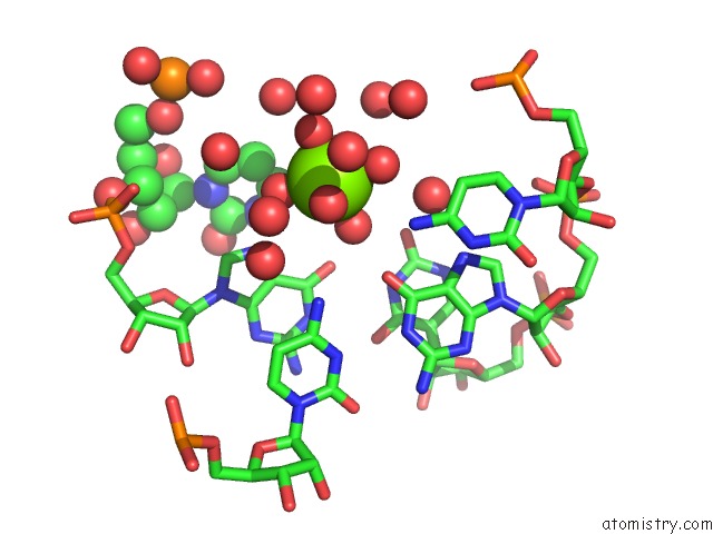
Mono view
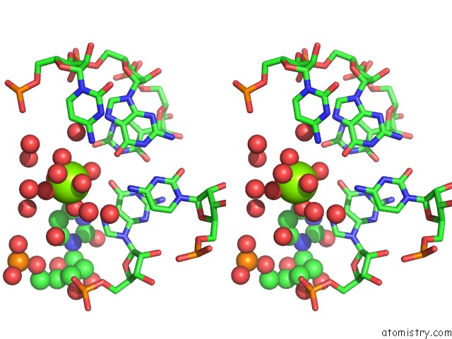
Stereo pair view
Mono view
Stereo pair view
A full contact list of Magnesium with other atoms in the Mg binding
site number 1 of X-Ray Structure Containing the Pseudouridylated U2 Snrna and Mammalian Intron Branch Site Consensus Sequences within 5.0Å range:
|
Reference:
Y.Lin,
C.L.Kielkopf.
X-Ray Structures of U2 Snrna-Branchpoint Duplexes Containing Conserved Pseudouridines. Biochemistry V. 47 5503 2008.
ISSN: ISSN 0006-2960
PubMed: 18435545
DOI: 10.1021/BI7022392
Page generated: Sun Aug 10 19:21:35 2025
ISSN: ISSN 0006-2960
PubMed: 18435545
DOI: 10.1021/BI7022392
Last articles
Mg in 4FO6Mg in 4FO0
Mg in 4FME
Mg in 4FMM
Mg in 4FNI
Mg in 4FMO
Mg in 4FMD
Mg in 4FMC
Mg in 4FMB
Mg in 4FM9