Magnesium »
PDB 3cfx-3cpj »
3chl »
Magnesium in PDB 3chl: Crystal Structure of Alpha-14 Giardin with Magnesium Bound
Protein crystallography data
The structure of Crystal Structure of Alpha-14 Giardin with Magnesium Bound, PDB code: 3chl
was solved by
P.Pathuri,
H.Luecke,
with X-Ray Crystallography technique. A brief refinement statistics is given in the table below:
| Resolution Low / High (Å) | 50.00 / 1.90 |
| Space group | H 3 |
| Cell size a, b, c (Å), α, β, γ (°) | 133.859, 133.859, 69.668, 90.00, 90.00, 120.00 |
| R / Rfree (%) | 19.9 / 23.1 |
Magnesium Binding Sites:
The binding sites of Magnesium atom in the Crystal Structure of Alpha-14 Giardin with Magnesium Bound
(pdb code 3chl). This binding sites where shown within
5.0 Angstroms radius around Magnesium atom.
In total only one binding site of Magnesium was determined in the Crystal Structure of Alpha-14 Giardin with Magnesium Bound, PDB code: 3chl:
In total only one binding site of Magnesium was determined in the Crystal Structure of Alpha-14 Giardin with Magnesium Bound, PDB code: 3chl:
Magnesium binding site 1 out of 1 in 3chl
Go back to
Magnesium binding site 1 out
of 1 in the Crystal Structure of Alpha-14 Giardin with Magnesium Bound
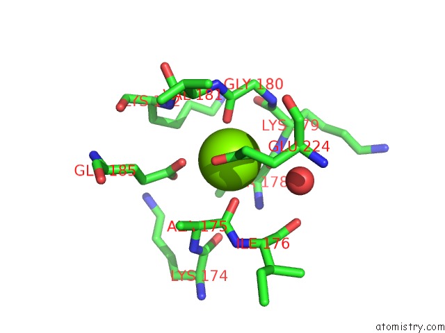
Mono view
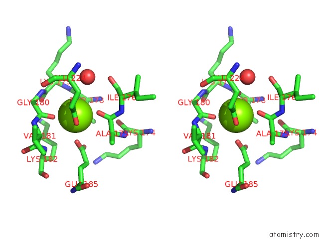
Stereo pair view
Mono view
Stereo pair view
A full contact list of Magnesium with other atoms in the Mg binding
site number 1 of Crystal Structure of Alpha-14 Giardin with Magnesium Bound within 5.0Å range:
|
Reference:
P.Pathuri,
E.T.Nguyen,
G.Ozorowski,
S.G.Svard,
H.Luecke.
Apo and Calcium-Bound Crystal Structures of Cytoskeletal Protein Alpha-14 Giardin (Annexin E1) From the Intestinal Protozoan Parasite Giardia Lamblia J.Mol.Biol. V. 385 1098 2009.
ISSN: ISSN 0022-2836
PubMed: 19046974
DOI: 10.1016/J.JMB.2008.11.012
Page generated: Sun Aug 10 19:21:48 2025
ISSN: ISSN 0022-2836
PubMed: 19046974
DOI: 10.1016/J.JMB.2008.11.012
Last articles
Mg in 4CZ1Mg in 4CYQ
Mg in 4CYP
Mg in 4CYO
Mg in 4CYJ
Mg in 4CW7
Mg in 4CYN
Mg in 4CYM
Mg in 4CWB
Mg in 4CVN