Magnesium »
PDB 3cfx-3cpj »
3ci4 »
Magnesium in PDB 3ci4: Structure of the Pduo-Type Atp:Co(I)Rrinoid Adenosyltransferase From Lactobacillus Reuteri Complexed with Four-Coordinate Cob(II)Inamide and Atp
Enzymatic activity of Structure of the Pduo-Type Atp:Co(I)Rrinoid Adenosyltransferase From Lactobacillus Reuteri Complexed with Four-Coordinate Cob(II)Inamide and Atp
All present enzymatic activity of Structure of the Pduo-Type Atp:Co(I)Rrinoid Adenosyltransferase From Lactobacillus Reuteri Complexed with Four-Coordinate Cob(II)Inamide and Atp:
2.5.1.17;
2.5.1.17;
Protein crystallography data
The structure of Structure of the Pduo-Type Atp:Co(I)Rrinoid Adenosyltransferase From Lactobacillus Reuteri Complexed with Four-Coordinate Cob(II)Inamide and Atp, PDB code: 3ci4
was solved by
M.St.Maurice,
P.E.Mera,
J.C.Escalante-Semerena,
I.Rayment,
with X-Ray Crystallography technique. A brief refinement statistics is given in the table below:
| Resolution Low / High (Å) | 30.00 / 2.00 |
| Space group | H 3 |
| Cell size a, b, c (Å), α, β, γ (°) | 67.826, 67.826, 111.260, 90.00, 90.00, 120.00 |
| R / Rfree (%) | 17 / 20.9 |
Other elements in 3ci4:
The structure of Structure of the Pduo-Type Atp:Co(I)Rrinoid Adenosyltransferase From Lactobacillus Reuteri Complexed with Four-Coordinate Cob(II)Inamide and Atp also contains other interesting chemical elements:
| Cobalt | (Co) | 1 atom |
| Potassium | (K) | 1 atom |
Magnesium Binding Sites:
The binding sites of Magnesium atom in the Structure of the Pduo-Type Atp:Co(I)Rrinoid Adenosyltransferase From Lactobacillus Reuteri Complexed with Four-Coordinate Cob(II)Inamide and Atp
(pdb code 3ci4). This binding sites where shown within
5.0 Angstroms radius around Magnesium atom.
In total only one binding site of Magnesium was determined in the Structure of the Pduo-Type Atp:Co(I)Rrinoid Adenosyltransferase From Lactobacillus Reuteri Complexed with Four-Coordinate Cob(II)Inamide and Atp, PDB code: 3ci4:
In total only one binding site of Magnesium was determined in the Structure of the Pduo-Type Atp:Co(I)Rrinoid Adenosyltransferase From Lactobacillus Reuteri Complexed with Four-Coordinate Cob(II)Inamide and Atp, PDB code: 3ci4:
Magnesium binding site 1 out of 1 in 3ci4
Go back to
Magnesium binding site 1 out
of 1 in the Structure of the Pduo-Type Atp:Co(I)Rrinoid Adenosyltransferase From Lactobacillus Reuteri Complexed with Four-Coordinate Cob(II)Inamide and Atp
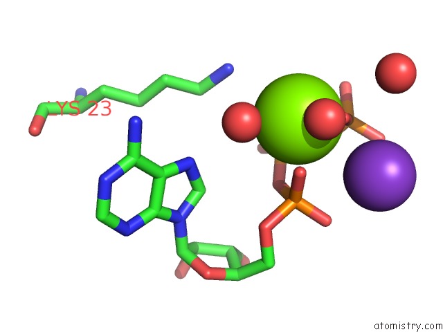
Mono view
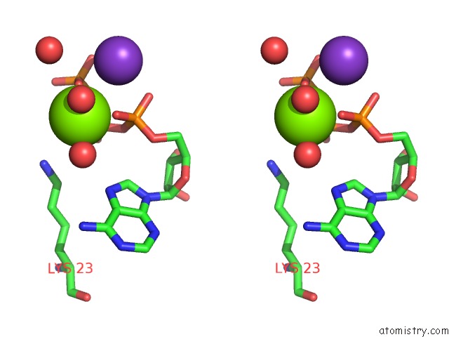
Stereo pair view
Mono view
Stereo pair view
A full contact list of Magnesium with other atoms in the Mg binding
site number 1 of Structure of the Pduo-Type Atp:Co(I)Rrinoid Adenosyltransferase From Lactobacillus Reuteri Complexed with Four-Coordinate Cob(II)Inamide and Atp within 5.0Å range:
|
Reference:
M.St Maurice,
P.Mera,
K.Park,
T.C.Brunold,
J.C.Escalante-Semerena,
I.Rayment.
Structural Characterization of A Human-Type Corrinoid Adenosyltransferase Confirms That Coenzyme B12 Is Synthesized Through A Four-Coordinate Intermediate. Biochemistry V. 47 5755 2008.
ISSN: ISSN 0006-2960
PubMed: 18452306
DOI: 10.1021/BI800132D
Page generated: Wed Aug 14 11:23:55 2024
ISSN: ISSN 0006-2960
PubMed: 18452306
DOI: 10.1021/BI800132D
Last articles
Cl in 7U68Cl in 7U8E
Cl in 7U55
Cl in 7U5Z
Cl in 7U4K
Cl in 7U34
Cl in 7U4J
Cl in 7U4H
Cl in 7U36
Cl in 7U33