Magnesium »
PDB 3cfx-3cpj »
3cif »
Magnesium in PDB 3cif: Crystal Structure of C153S Mutant Glyceraldehyde 3-Phosphate Dehydrogenase From Cryptosporidium Parvum
Enzymatic activity of Crystal Structure of C153S Mutant Glyceraldehyde 3-Phosphate Dehydrogenase From Cryptosporidium Parvum
All present enzymatic activity of Crystal Structure of C153S Mutant Glyceraldehyde 3-Phosphate Dehydrogenase From Cryptosporidium Parvum:
1.2.1.12;
1.2.1.12;
Protein crystallography data
The structure of Crystal Structure of C153S Mutant Glyceraldehyde 3-Phosphate Dehydrogenase From Cryptosporidium Parvum, PDB code: 3cif
was solved by
W.J.Cook,
O.Senkovich,
D.Chattopadhyay,
with X-Ray Crystallography technique. A brief refinement statistics is given in the table below:
| Resolution Low / High (Å) | 20.00 / 2.00 |
| Space group | P 1 21 1 |
| Cell size a, b, c (Å), α, β, γ (°) | 68.000, 120.100, 79.280, 90.00, 92.08, 90.00 |
| R / Rfree (%) | 17.8 / 21 |
Magnesium Binding Sites:
The binding sites of Magnesium atom in the Crystal Structure of C153S Mutant Glyceraldehyde 3-Phosphate Dehydrogenase From Cryptosporidium Parvum
(pdb code 3cif). This binding sites where shown within
5.0 Angstroms radius around Magnesium atom.
In total 2 binding sites of Magnesium where determined in the Crystal Structure of C153S Mutant Glyceraldehyde 3-Phosphate Dehydrogenase From Cryptosporidium Parvum, PDB code: 3cif:
Jump to Magnesium binding site number: 1; 2;
In total 2 binding sites of Magnesium where determined in the Crystal Structure of C153S Mutant Glyceraldehyde 3-Phosphate Dehydrogenase From Cryptosporidium Parvum, PDB code: 3cif:
Jump to Magnesium binding site number: 1; 2;
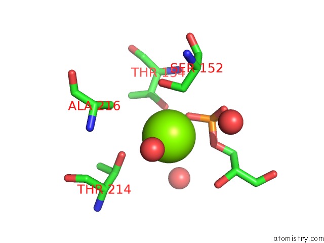
Magnesium binding site 1 out of 2 in 3cif
Go back to
Magnesium binding site 1 out
of 2 in the Crystal Structure of C153S Mutant Glyceraldehyde 3-Phosphate Dehydrogenase From Cryptosporidium Parvum

Mono view

Stereo pair view
Mono view
Stereo pair view
A full contact list of Magnesium with other atoms in the Mg binding
site number 1 of Crystal Structure of C153S Mutant Glyceraldehyde 3-Phosphate Dehydrogenase From Cryptosporidium Parvum within 5.0Å range:
|
Magnesium binding site 2 out of 2 in 3cif
Go back to
Magnesium binding site 2 out
of 2 in the Crystal Structure of C153S Mutant Glyceraldehyde 3-Phosphate Dehydrogenase From Cryptosporidium Parvum
Mono view

Stereo pair view
Mono view
Stereo pair view
A full contact list of Magnesium with other atoms in the Mg binding
site number 2 of Crystal Structure of C153S Mutant Glyceraldehyde 3-Phosphate Dehydrogenase From Cryptosporidium Parvum within 5.0Å range:
|
Reference:
W.J.Cook,
O.Senkovich,
D.Chattopadhyay.
An Unexpected Phosphate Binding Site in Glyceraldehyde 3-Phosphate Dehydrogenase: Crystal Structures of Apo, Holo and Ternary Complex of Cryptosporidium Parvum Enzyme Bmc Struct.Biol. V. 9 9 2009.
ISSN: ESSN 1472-6807
PubMed: 19243605
DOI: 10.1186/1472-6807-9-9
Page generated: Sun Aug 10 19:22:18 2025
ISSN: ESSN 1472-6807
PubMed: 19243605
DOI: 10.1186/1472-6807-9-9
Last articles
Mg in 5MMJMg in 5MRA
Mg in 5MTV
Mg in 5MS0
Mg in 5MRU
Mg in 5MQJ
Mg in 5MQW
Mg in 5MQT
Mg in 5MQL
Mg in 5MQ1