Magnesium »
PDB 3cpw-3d19 »
3crc »
Magnesium in PDB 3crc: Crystal Structure of Escherichia Coli Mazg, the Regulator of Nutritional Stress Response
Protein crystallography data
The structure of Crystal Structure of Escherichia Coli Mazg, the Regulator of Nutritional Stress Response, PDB code: 3crc
was solved by
S.Lee,
M.H.Kim,
B.S.Kang,
J.S.Kim,
Y.G.Kim,
K.J.Kim,
with X-Ray Crystallography technique. A brief refinement statistics is given in the table below:
| Resolution Low / High (Å) | 33.79 / 3.00 |
| Space group | P 21 21 21 |
| Cell size a, b, c (Å), α, β, γ (°) | 63.230, 66.910, 140.700, 90.00, 90.00, 90.00 |
| R / Rfree (%) | 21.6 / 34.5 |
Magnesium Binding Sites:
The binding sites of Magnesium atom in the Crystal Structure of Escherichia Coli Mazg, the Regulator of Nutritional Stress Response
(pdb code 3crc). This binding sites where shown within
5.0 Angstroms radius around Magnesium atom.
In total only one binding site of Magnesium was determined in the Crystal Structure of Escherichia Coli Mazg, the Regulator of Nutritional Stress Response, PDB code: 3crc:
In total only one binding site of Magnesium was determined in the Crystal Structure of Escherichia Coli Mazg, the Regulator of Nutritional Stress Response, PDB code: 3crc:
Magnesium binding site 1 out of 1 in 3crc
Go back to
Magnesium binding site 1 out
of 1 in the Crystal Structure of Escherichia Coli Mazg, the Regulator of Nutritional Stress Response
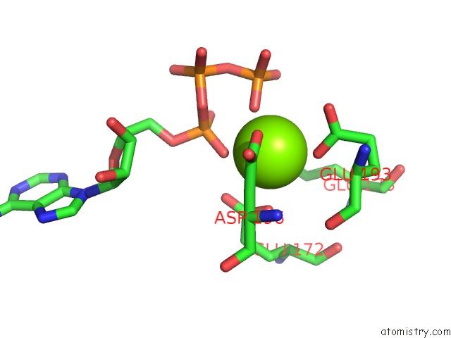
Mono view
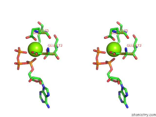
Stereo pair view
Mono view
Stereo pair view
A full contact list of Magnesium with other atoms in the Mg binding
site number 1 of Crystal Structure of Escherichia Coli Mazg, the Regulator of Nutritional Stress Response within 5.0Å range:
|
Reference:
S.Lee,
M.H.Kim,
B.S.Kang,
J.S.Kim,
G.H.Kim,
Y.G.Kim,
K.J.Kim.
Crystal Structure of Escherichia Coli Mazg, the Regulator of Nutritional Stress Response. J.Biol.Chem. V. 283 15232 2008.
ISSN: ISSN 0021-9258
PubMed: 18353782
DOI: 10.1074/JBC.M800479200
Page generated: Sun Aug 10 19:41:01 2025
ISSN: ISSN 0021-9258
PubMed: 18353782
DOI: 10.1074/JBC.M800479200
Last articles
Mg in 3ZKDMg in 3ZLB
Mg in 3ZLM
Mg in 3ZIA
Mg in 3ZL6
Mg in 3ZJC
Mg in 3ZJY
Mg in 3ZJT
Mg in 3ZJU
Mg in 3ZHV