Magnesium »
PDB 3cpw-3d19 »
3csk »
Magnesium in PDB 3csk: Structure of Dpp III From Saccharomyces Cerevisiae
Enzymatic activity of Structure of Dpp III From Saccharomyces Cerevisiae
All present enzymatic activity of Structure of Dpp III From Saccharomyces Cerevisiae:
3.4.14.4;
3.4.14.4;
Protein crystallography data
The structure of Structure of Dpp III From Saccharomyces Cerevisiae, PDB code: 3csk
was solved by
P.K.Baral,
N.Jajcanin,
S.Deller,
P.Macheroux,
M.Abramic,
K.Gruber,
with X-Ray Crystallography technique. A brief refinement statistics is given in the table below:
| Resolution Low / High (Å) | 30.00 / 1.95 |
| Space group | P 1 21 1 |
| Cell size a, b, c (Å), α, β, γ (°) | 60.621, 110.121, 67.908, 90.00, 113.45, 90.00 |
| R / Rfree (%) | 19.2 / 22.8 |
Other elements in 3csk:
The structure of Structure of Dpp III From Saccharomyces Cerevisiae also contains other interesting chemical elements:
| Zinc | (Zn) | 1 atom |
Magnesium Binding Sites:
The binding sites of Magnesium atom in the Structure of Dpp III From Saccharomyces Cerevisiae
(pdb code 3csk). This binding sites where shown within
5.0 Angstroms radius around Magnesium atom.
In total 2 binding sites of Magnesium where determined in the Structure of Dpp III From Saccharomyces Cerevisiae, PDB code: 3csk:
Jump to Magnesium binding site number: 1; 2;
In total 2 binding sites of Magnesium where determined in the Structure of Dpp III From Saccharomyces Cerevisiae, PDB code: 3csk:
Jump to Magnesium binding site number: 1; 2;
Magnesium binding site 1 out of 2 in 3csk
Go back to
Magnesium binding site 1 out
of 2 in the Structure of Dpp III From Saccharomyces Cerevisiae
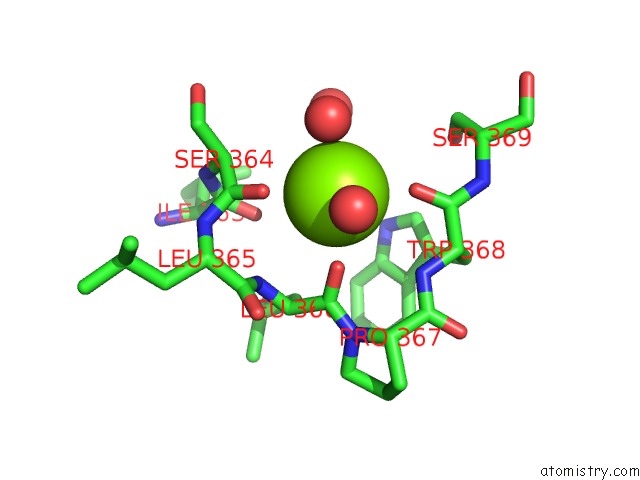
Mono view
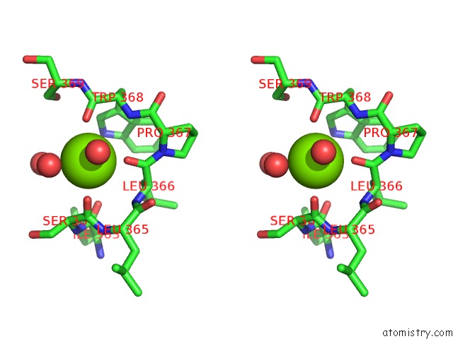
Stereo pair view
Mono view
Stereo pair view
A full contact list of Magnesium with other atoms in the Mg binding
site number 1 of Structure of Dpp III From Saccharomyces Cerevisiae within 5.0Å range:
|
Magnesium binding site 2 out of 2 in 3csk
Go back to
Magnesium binding site 2 out
of 2 in the Structure of Dpp III From Saccharomyces Cerevisiae
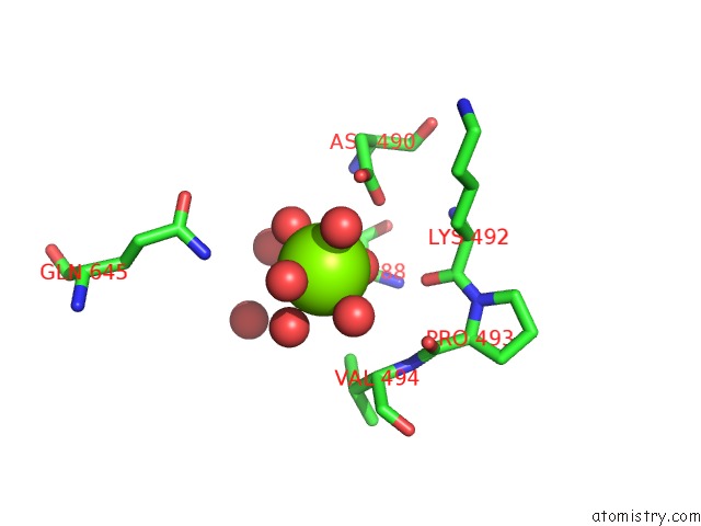
Mono view
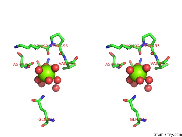
Stereo pair view
Mono view
Stereo pair view
A full contact list of Magnesium with other atoms in the Mg binding
site number 2 of Structure of Dpp III From Saccharomyces Cerevisiae within 5.0Å range:
|
Reference:
P.K.Baral,
N.Jajcanin-Jozic,
S.Deller,
P.Macheroux,
M.Abramic,
K.Gruber.
The First Structure of Dipeptidyl-Peptidase III Provides Insight Into the Catalytic Mechanism and Mode of Substrate Binding. J.Biol.Chem. V. 283 22316 2008.
ISSN: ISSN 0021-9258
PubMed: 18550518
DOI: 10.1074/JBC.M803522200
Page generated: Wed Aug 14 11:52:08 2024
ISSN: ISSN 0021-9258
PubMed: 18550518
DOI: 10.1074/JBC.M803522200
Last articles
Fe in 2YXOFe in 2YRS
Fe in 2YXC
Fe in 2YNM
Fe in 2YVJ
Fe in 2YP1
Fe in 2YU2
Fe in 2YU1
Fe in 2YQB
Fe in 2YOO