Magnesium »
PDB 3cpw-3d19 »
3cur »
Magnesium in PDB 3cur: Structure of A Double Methionine Mutant of Ni-Fe Hydrogenase
Enzymatic activity of Structure of A Double Methionine Mutant of Ni-Fe Hydrogenase
All present enzymatic activity of Structure of A Double Methionine Mutant of Ni-Fe Hydrogenase:
1.12.2.1;
1.12.2.1;
Protein crystallography data
The structure of Structure of A Double Methionine Mutant of Ni-Fe Hydrogenase, PDB code: 3cur
was solved by
A.Volbeda,
with X-Ray Crystallography technique. A brief refinement statistics is given in the table below:
| Resolution Low / High (Å) | 24.99 / 2.40 |
| Space group | P 1 21 1 |
| Cell size a, b, c (Å), α, β, γ (°) | 64.600, 99.900, 183.000, 90.00, 91.60, 90.00 |
| R / Rfree (%) | 15 / 19.4 |
Other elements in 3cur:
The structure of Structure of A Double Methionine Mutant of Ni-Fe Hydrogenase also contains other interesting chemical elements:
| Nickel | (Ni) | 3 atoms |
| Iron | (Fe) | 36 atoms |
Magnesium Binding Sites:
The binding sites of Magnesium atom in the Structure of A Double Methionine Mutant of Ni-Fe Hydrogenase
(pdb code 3cur). This binding sites where shown within
5.0 Angstroms radius around Magnesium atom.
In total 3 binding sites of Magnesium where determined in the Structure of A Double Methionine Mutant of Ni-Fe Hydrogenase, PDB code: 3cur:
Jump to Magnesium binding site number: 1; 2; 3;
In total 3 binding sites of Magnesium where determined in the Structure of A Double Methionine Mutant of Ni-Fe Hydrogenase, PDB code: 3cur:
Jump to Magnesium binding site number: 1; 2; 3;
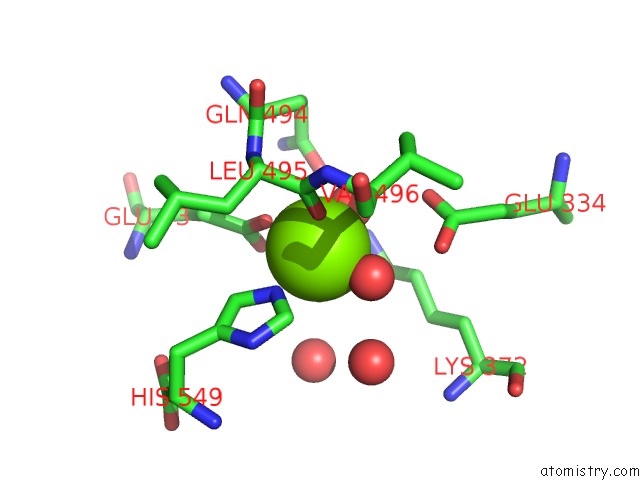
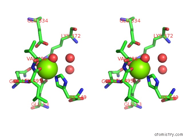
Magnesium binding site 1 out of 3 in 3cur
Go back to
Magnesium binding site 1 out
of 3 in the Structure of A Double Methionine Mutant of Ni-Fe Hydrogenase
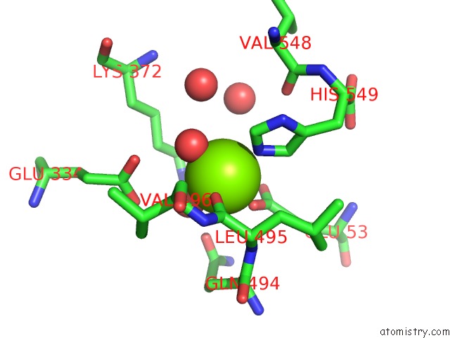
Mono view
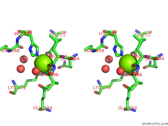
Stereo pair view
Mono view
Stereo pair view
A full contact list of Magnesium with other atoms in the Mg binding
site number 1 of Structure of A Double Methionine Mutant of Ni-Fe Hydrogenase within 5.0Å range:
|
Magnesium binding site 2 out of 3 in 3cur
Go back to
Magnesium binding site 2 out
of 3 in the Structure of A Double Methionine Mutant of Ni-Fe Hydrogenase
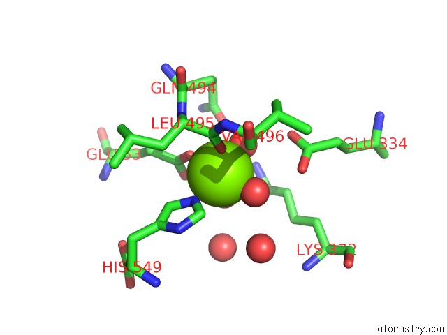
Mono view
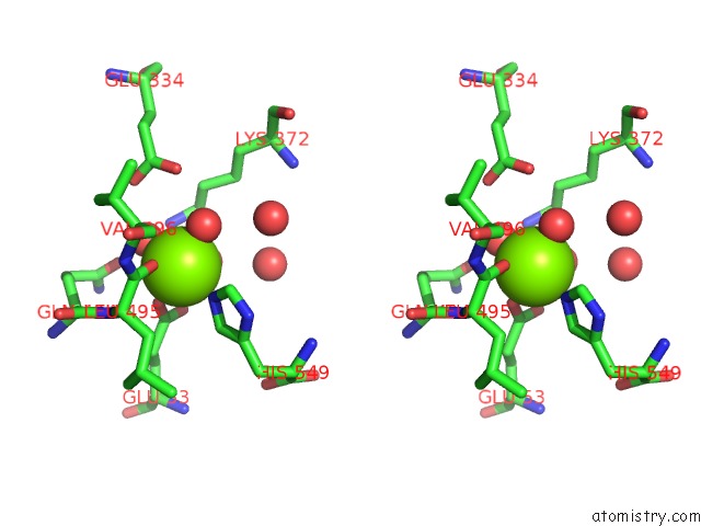
Stereo pair view
Mono view
Stereo pair view
A full contact list of Magnesium with other atoms in the Mg binding
site number 2 of Structure of A Double Methionine Mutant of Ni-Fe Hydrogenase within 5.0Å range:
|
Magnesium binding site 3 out of 3 in 3cur
Go back to
Magnesium binding site 3 out
of 3 in the Structure of A Double Methionine Mutant of Ni-Fe Hydrogenase
Mono view
Stereo pair view
Mono view
Stereo pair view
A full contact list of Magnesium with other atoms in the Mg binding
site number 3 of Structure of A Double Methionine Mutant of Ni-Fe Hydrogenase within 5.0Å range:
|
Reference:
F.Leroux,
S.Dementin,
B.Burlat,
L.Cournac,
A.Volbeda,
S.Champ,
L.Martin,
B.Guigliarelli,
P.Bertrand,
J.Fontecilla-Camps,
M.Rousset.
Experimental Approaches to Kinetics of Gas Diffusion in Hydrogenase Proc.Natl.Acad.Sci.Usa V. 105 11188 2008.
ISSN: ISSN 0027-8424
PubMed: 18685111
DOI: 10.1073/PNAS.0803689105
Page generated: Wed Aug 14 11:53:59 2024
ISSN: ISSN 0027-8424
PubMed: 18685111
DOI: 10.1073/PNAS.0803689105
Last articles
Zn in 9MJ5Zn in 9HNW
Zn in 9G0L
Zn in 9FNE
Zn in 9DZN
Zn in 9E0I
Zn in 9D32
Zn in 9DAK
Zn in 8ZXC
Zn in 8ZUF