Magnesium »
PDB 3cpw-3d19 »
3cyi »
Magnesium in PDB 3cyi: Crystal Structure of Human Sulfiredoxin (Srx) in Complex with Atp:MG2+
Enzymatic activity of Crystal Structure of Human Sulfiredoxin (Srx) in Complex with Atp:MG2+
All present enzymatic activity of Crystal Structure of Human Sulfiredoxin (Srx) in Complex with Atp:MG2+:
1.8.98.2;
1.8.98.2;
Protein crystallography data
The structure of Crystal Structure of Human Sulfiredoxin (Srx) in Complex with Atp:MG2+, PDB code: 3cyi
was solved by
T.J.Jonsson,
M.S.Murray,
L.C.Johnson,
W.T.Lowther,
with X-Ray Crystallography technique. A brief refinement statistics is given in the table below:
| Resolution Low / High (Å) | 38.70 / 1.80 |
| Space group | P 32 2 1 |
| Cell size a, b, c (Å), α, β, γ (°) | 68.530, 68.530, 51.090, 90.00, 90.00, 120.00 |
| R / Rfree (%) | 20.1 / 23.1 |
Magnesium Binding Sites:
The binding sites of Magnesium atom in the Crystal Structure of Human Sulfiredoxin (Srx) in Complex with Atp:MG2+
(pdb code 3cyi). This binding sites where shown within
5.0 Angstroms radius around Magnesium atom.
In total only one binding site of Magnesium was determined in the Crystal Structure of Human Sulfiredoxin (Srx) in Complex with Atp:MG2+, PDB code: 3cyi:
In total only one binding site of Magnesium was determined in the Crystal Structure of Human Sulfiredoxin (Srx) in Complex with Atp:MG2+, PDB code: 3cyi:
Magnesium binding site 1 out of 1 in 3cyi
Go back to
Magnesium binding site 1 out
of 1 in the Crystal Structure of Human Sulfiredoxin (Srx) in Complex with Atp:MG2+
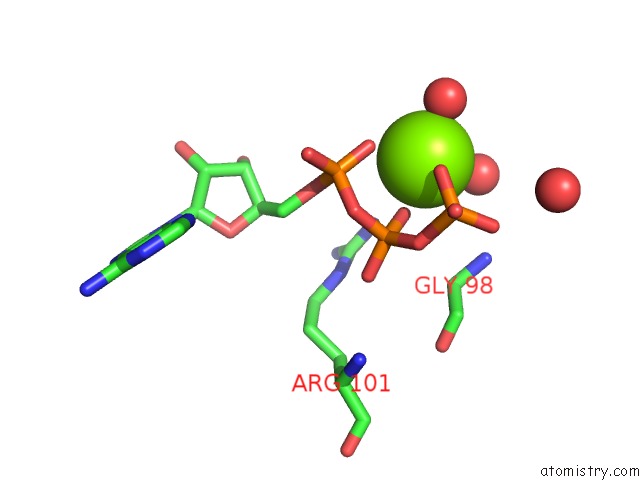
Mono view
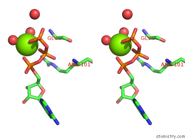
Stereo pair view
Mono view
Stereo pair view
A full contact list of Magnesium with other atoms in the Mg binding
site number 1 of Crystal Structure of Human Sulfiredoxin (Srx) in Complex with Atp:MG2+ within 5.0Å range:
|
Reference:
T.J.Jonsson,
M.S.Murray,
L.C.Johnson,
W.T.Lowther.
Reduction of Cysteine Sulfinic Acid in Peroxiredoxin By Sulfiredoxin Proceeds Directly Through A Sulfinic Phosphoryl Ester Intermediate. J.Biol.Chem. V. 283 23846 2008.
ISSN: ISSN 0021-9258
PubMed: 18579529
DOI: 10.1074/JBC.M803244200
Page generated: Sun Aug 10 19:46:14 2025
ISSN: ISSN 0021-9258
PubMed: 18579529
DOI: 10.1074/JBC.M803244200
Last articles
Mg in 6LU1Mg in 6LT4
Mg in 6LY7
Mg in 6LY6
Mg in 6LY3
Mg in 6LX1
Mg in 6LTW
Mg in 6LVW
Mg in 6LUH
Mg in 6LTS