Magnesium in PDB 3d47: Crystal Structure of L-Rhamnonate Dehydratase From Salmonella Typhimurium Complexed with Mg and D-Malate
Protein crystallography data
The structure of Crystal Structure of L-Rhamnonate Dehydratase From Salmonella Typhimurium Complexed with Mg and D-Malate, PDB code: 3d47
was solved by
A.A.Fedorov,
E.V.Fedorov,
J.M.Sauder,
S.K.Burley,
J.A.Gerlt,
S.C.Almo,
Newyork Sgx Research Center For Structural Genomics (Nysgxrc),
with X-Ray Crystallography technique. A brief refinement statistics is given in the table below:
Resolution Low / High (Å)
|
24.88 /
1.80
|
Space group
|
P 1 21 1
|
Cell size a, b, c (Å), α, β, γ (°)
|
98.222,
122.003,
141.350,
90.00,
108.24,
90.00
|
R / Rfree (%)
|
20.2 /
23.2
|
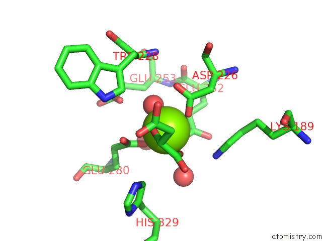
Magnesium Binding Sites:
The binding sites of Magnesium atom in the Crystal Structure of L-Rhamnonate Dehydratase From Salmonella Typhimurium Complexed with Mg and D-Malate
(pdb code 3d47). This binding sites where shown within
5.0 Angstroms radius around Magnesium atom.
In total 8 binding sites of Magnesium where determined in the
Crystal Structure of L-Rhamnonate Dehydratase From Salmonella Typhimurium Complexed with Mg and D-Malate, PDB code: 3d47:
Jump to Magnesium binding site number:
1;
2;
3;
4;
5;
6;
7;
8;
Magnesium binding site 1 out
of 8 in 3d47
Go back to
Magnesium Binding Sites List in 3d47
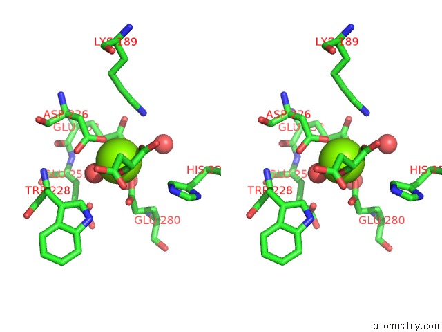
Magnesium binding site 1 out
of 8 in the Crystal Structure of L-Rhamnonate Dehydratase From Salmonella Typhimurium Complexed with Mg and D-Malate
 Mono view
 Stereo pair view
|
A full contact list of Magnesium with other atoms in the Mg binding
site number 1 of Crystal Structure of L-Rhamnonate Dehydratase From Salmonella Typhimurium Complexed with Mg and D-Malate within 5.0Å range:
probe
|
atom
|
residue
|
distance (Å)
|
B
|
Occ
|
A:Mg501
b:12.8
occ:1.00
|
OE1
|
A:GLU280
|
2.0
|
12.1
|
1.0
|
OD2
|
A:ASP226
|
2.0
|
10.8
|
1.0
|
O
|
A:HOH551
|
2.1
|
12.2
|
1.0
|
O3
|
A:MLT502
|
2.1
|
12.8
|
1.0
|
OE1
|
A:GLU252
|
2.2
|
9.2
|
1.0
|
O1
|
A:MLT502
|
2.2
|
14.1
|
1.0
|
CD
|
A:GLU280
|
2.9
|
12.7
|
1.0
|
C1
|
A:MLT502
|
2.9
|
16.7
|
1.0
|
C2
|
A:MLT502
|
3.0
|
16.4
|
1.0
|
CG
|
A:ASP226
|
3.1
|
10.9
|
1.0
|
CD
|
A:GLU252
|
3.1
|
10.3
|
1.0
|
OE2
|
A:GLU280
|
3.2
|
13.4
|
1.0
|
OD1
|
A:ASP226
|
3.4
|
11.3
|
1.0
|
CG
|
A:GLU252
|
3.8
|
8.1
|
1.0
|
O
|
A:HOH520
|
3.9
|
13.4
|
1.0
|
NZ
|
A:LYS189
|
4.0
|
13.5
|
1.0
|
C3
|
A:MLT502
|
4.0
|
19.0
|
1.0
|
OE2
|
A:GLU252
|
4.1
|
11.7
|
1.0
|
O2
|
A:MLT502
|
4.1
|
15.3
|
1.0
|
O
|
A:HOH522
|
4.2
|
13.3
|
1.0
|
OE1
|
A:GLU253
|
4.2
|
13.4
|
1.0
|
CG
|
A:GLU280
|
4.3
|
13.3
|
1.0
|
CD2
|
A:HIS329
|
4.3
|
21.2
|
1.0
|
CB
|
A:ASP226
|
4.4
|
9.4
|
1.0
|
CE
|
A:LYS189
|
4.6
|
12.9
|
1.0
|
CB
|
A:GLU253
|
4.9
|
9.8
|
1.0
|
CD1
|
A:TRP228
|
5.0
|
12.0
|
1.0
|
CB
|
A:GLU280
|
5.0
|
13.0
|
1.0
|
NE2
|
A:HIS329
|
5.0
|
21.4
|
1.0
|
|
Magnesium binding site 2 out
of 8 in 3d47
Go back to
Magnesium Binding Sites List in 3d47
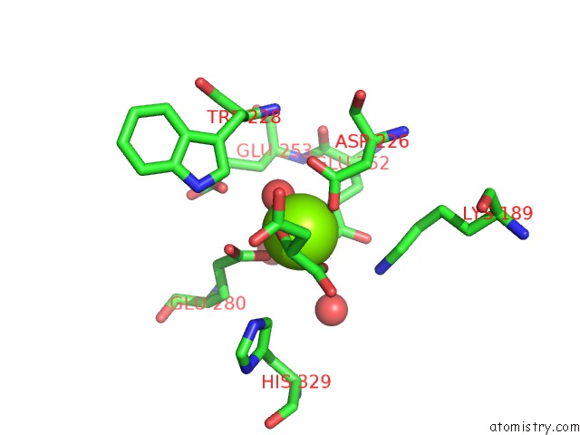
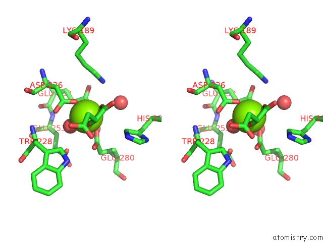
Magnesium binding site 2 out
of 8 in the Crystal Structure of L-Rhamnonate Dehydratase From Salmonella Typhimurium Complexed with Mg and D-Malate
 Mono view
 Stereo pair view
|
A full contact list of Magnesium with other atoms in the Mg binding
site number 2 of Crystal Structure of L-Rhamnonate Dehydratase From Salmonella Typhimurium Complexed with Mg and D-Malate within 5.0Å range:
probe
|
atom
|
residue
|
distance (Å)
|
B
|
Occ
|
B:Mg501
b:17.5
occ:1.00
|
O
|
B:HOH506
|
2.0
|
17.4
|
1.0
|
OD2
|
B:ASP226
|
2.0
|
16.8
|
1.0
|
OE1
|
B:GLU252
|
2.0
|
13.9
|
1.0
|
OE1
|
B:GLU280
|
2.0
|
17.4
|
1.0
|
O3
|
B:MLT502
|
2.1
|
21.8
|
1.0
|
O1
|
B:MLT502
|
2.1
|
22.2
|
1.0
|
C1
|
B:MLT502
|
2.9
|
25.0
|
1.0
|
C2
|
B:MLT502
|
3.0
|
24.7
|
1.0
|
CG
|
B:ASP226
|
3.0
|
16.1
|
1.0
|
CD
|
B:GLU280
|
3.0
|
17.3
|
1.0
|
CD
|
B:GLU252
|
3.0
|
16.6
|
1.0
|
OE2
|
B:GLU280
|
3.3
|
17.4
|
1.0
|
OD1
|
B:ASP226
|
3.3
|
16.1
|
1.0
|
CG
|
B:GLU252
|
3.7
|
16.0
|
1.0
|
OE2
|
B:GLU252
|
3.9
|
14.6
|
1.0
|
NZ
|
B:LYS189
|
4.0
|
19.4
|
1.0
|
C3
|
B:MLT502
|
4.1
|
27.6
|
1.0
|
O2
|
B:MLT502
|
4.1
|
24.3
|
1.0
|
O
|
B:HOH590
|
4.1
|
19.4
|
1.0
|
O
|
B:HOH515
|
4.2
|
19.9
|
1.0
|
OE1
|
B:GLU253
|
4.3
|
19.0
|
1.0
|
CG
|
B:GLU280
|
4.3
|
17.6
|
1.0
|
CB
|
B:ASP226
|
4.3
|
16.7
|
1.0
|
CD2
|
B:HIS329
|
4.4
|
26.4
|
1.0
|
CE
|
B:LYS189
|
4.6
|
18.8
|
1.0
|
CB
|
B:GLU252
|
4.9
|
18.8
|
1.0
|
CD1
|
B:TRP228
|
5.0
|
14.8
|
1.0
|
CB
|
B:GLU280
|
5.0
|
16.0
|
1.0
|
|
Magnesium binding site 3 out
of 8 in 3d47
Go back to
Magnesium Binding Sites List in 3d47
Magnesium binding site 3 out
of 8 in the Crystal Structure of L-Rhamnonate Dehydratase From Salmonella Typhimurium Complexed with Mg and D-Malate
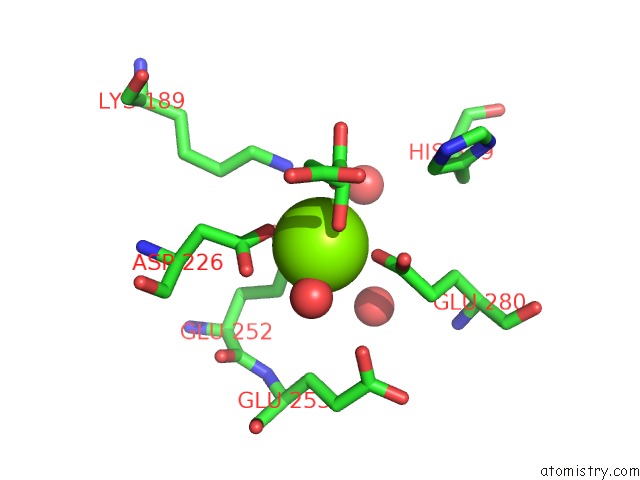
 Mono view
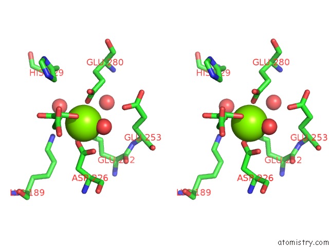
 Stereo pair view
|
A full contact list of Magnesium with other atoms in the Mg binding
site number 3 of Crystal Structure of L-Rhamnonate Dehydratase From Salmonella Typhimurium Complexed with Mg and D-Malate within 5.0Å range:
probe
|
atom
|
residue
|
distance (Å)
|
B
|
Occ
|
C:Mg501
b:15.0
occ:1.00
|
OE1
|
C:GLU252
|
1.9
|
8.7
|
1.0
|
OD2
|
C:ASP226
|
2.0
|
10.3
|
1.0
|
O
|
C:HOH517
|
2.1
|
12.6
|
1.0
|
OE1
|
C:GLU280
|
2.1
|
13.4
|
1.0
|
O1
|
C:MLT502
|
2.3
|
16.8
|
1.0
|
O3
|
C:MLT502
|
2.3
|
17.5
|
1.0
|
CD
|
C:GLU252
|
2.9
|
12.9
|
1.0
|
CG
|
C:ASP226
|
3.0
|
10.8
|
1.0
|
CD
|
C:GLU280
|
3.1
|
16.9
|
1.0
|
C1
|
C:MLT502
|
3.1
|
19.2
|
1.0
|
C2
|
C:MLT502
|
3.2
|
19.1
|
1.0
|
OD1
|
C:ASP226
|
3.3
|
12.8
|
1.0
|
OE2
|
C:GLU280
|
3.5
|
17.6
|
1.0
|
CG
|
C:GLU252
|
3.5
|
11.6
|
1.0
|
OE2
|
C:GLU252
|
3.8
|
11.0
|
1.0
|
NZ
|
C:LYS189
|
3.9
|
14.3
|
1.0
|
O
|
C:HOH530
|
4.0
|
15.7
|
1.0
|
O
|
C:HOH539
|
4.1
|
19.1
|
1.0
|
C3
|
C:MLT502
|
4.2
|
22.1
|
1.0
|
OE1
|
C:GLU253
|
4.2
|
13.1
|
1.0
|
O2
|
C:MLT502
|
4.3
|
19.4
|
1.0
|
CB
|
C:ASP226
|
4.3
|
10.7
|
1.0
|
CG
|
C:GLU280
|
4.3
|
16.1
|
1.0
|
CE
|
C:LYS189
|
4.5
|
14.8
|
1.0
|
CB
|
C:GLU252
|
4.7
|
11.9
|
1.0
|
CB
|
C:GLU253
|
4.9
|
14.7
|
1.0
|
CD2
|
C:HIS329
|
4.9
|
24.8
|
1.0
|
|
Magnesium binding site 4 out
of 8 in 3d47
Go back to
Magnesium Binding Sites List in 3d47
Magnesium binding site 4 out
of 8 in the Crystal Structure of L-Rhamnonate Dehydratase From Salmonella Typhimurium Complexed with Mg and D-Malate
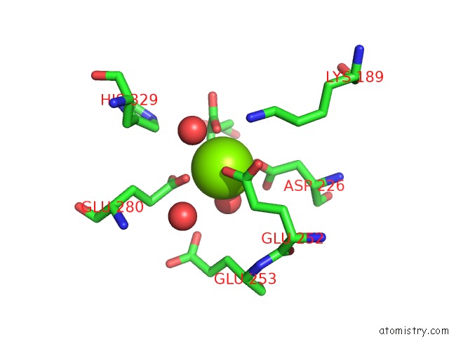
 Mono view
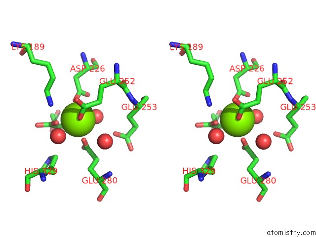
 Stereo pair view
|
A full contact list of Magnesium with other atoms in the Mg binding
site number 4 of Crystal Structure of L-Rhamnonate Dehydratase From Salmonella Typhimurium Complexed with Mg and D-Malate within 5.0Å range:
probe
|
atom
|
residue
|
distance (Å)
|
B
|
Occ
|
D:Mg501
b:18.4
occ:1.00
|
OE1
|
D:GLU252
|
2.0
|
18.7
|
1.0
|
OD2
|
D:ASP226
|
2.0
|
20.2
|
1.0
|
OE1
|
D:GLU280
|
2.1
|
19.1
|
1.0
|
O1
|
D:MLT502
|
2.2
|
26.9
|
1.0
|
O
|
D:HOH504
|
2.2
|
14.8
|
1.0
|
O3
|
D:MLT502
|
2.4
|
27.1
|
1.0
|
CD
|
D:GLU252
|
2.9
|
21.0
|
1.0
|
C1
|
D:MLT502
|
3.0
|
29.4
|
1.0
|
CG
|
D:ASP226
|
3.1
|
19.9
|
1.0
|
CD
|
D:GLU280
|
3.1
|
20.9
|
1.0
|
C2
|
D:MLT502
|
3.2
|
28.9
|
1.0
|
OD1
|
D:ASP226
|
3.5
|
19.9
|
1.0
|
OE2
|
D:GLU280
|
3.5
|
21.1
|
1.0
|
CG
|
D:GLU252
|
3.5
|
19.2
|
1.0
|
NZ
|
D:LYS189
|
3.7
|
17.0
|
1.0
|
OE2
|
D:GLU252
|
3.8
|
20.3
|
1.0
|
O
|
D:HOH518
|
3.9
|
21.6
|
1.0
|
O
|
D:HOH509
|
4.0
|
16.6
|
1.0
|
O2
|
D:MLT502
|
4.2
|
27.6
|
1.0
|
C3
|
D:MLT502
|
4.2
|
31.0
|
1.0
|
CB
|
D:ASP226
|
4.3
|
19.8
|
1.0
|
CD2
|
D:HIS329
|
4.4
|
27.5
|
1.0
|
CE
|
D:LYS189
|
4.4
|
18.6
|
1.0
|
OE1
|
D:GLU253
|
4.4
|
18.4
|
1.0
|
CG
|
D:GLU280
|
4.4
|
18.4
|
1.0
|
CB
|
D:GLU252
|
4.8
|
17.8
|
1.0
|
|
Magnesium binding site 5 out
of 8 in 3d47
Go back to
Magnesium Binding Sites List in 3d47
Magnesium binding site 5 out
of 8 in the Crystal Structure of L-Rhamnonate Dehydratase From Salmonella Typhimurium Complexed with Mg and D-Malate
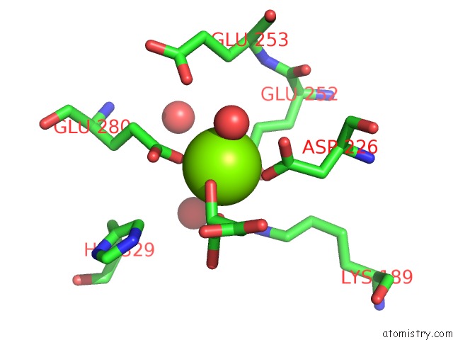
 Mono view
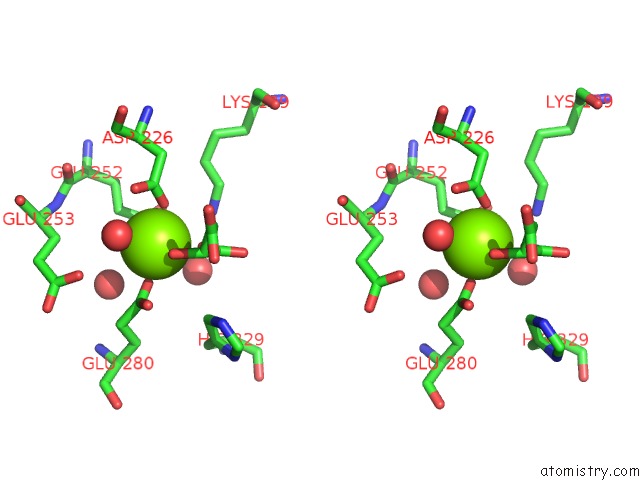
 Stereo pair view
|
A full contact list of Magnesium with other atoms in the Mg binding
site number 5 of Crystal Structure of L-Rhamnonate Dehydratase From Salmonella Typhimurium Complexed with Mg and D-Malate within 5.0Å range:
probe
|
atom
|
residue
|
distance (Å)
|
B
|
Occ
|
E:Mg501
b:20.8
occ:1.00
|
OE1
|
E:GLU280
|
1.9
|
20.3
|
1.0
|
OD2
|
E:ASP226
|
2.0
|
21.4
|
1.0
|
OE1
|
E:GLU252
|
2.1
|
14.3
|
1.0
|
O
|
E:HOH541
|
2.1
|
16.4
|
1.0
|
O3
|
E:MLT502
|
2.2
|
26.9
|
1.0
|
O1
|
E:MLT502
|
2.3
|
26.8
|
1.0
|
CD
|
E:GLU280
|
2.9
|
20.5
|
1.0
|
CD
|
E:GLU252
|
3.0
|
15.3
|
1.0
|
CG
|
E:ASP226
|
3.0
|
21.2
|
1.0
|
C1
|
E:MLT502
|
3.1
|
28.0
|
1.0
|
C2
|
E:MLT502
|
3.1
|
28.2
|
1.0
|
OE2
|
E:GLU280
|
3.4
|
18.7
|
1.0
|
OD1
|
E:ASP226
|
3.4
|
20.1
|
1.0
|
CG
|
E:GLU252
|
3.6
|
15.1
|
1.0
|
O
|
E:HOH515
|
3.9
|
13.8
|
1.0
|
OE2
|
E:GLU252
|
4.0
|
17.6
|
1.0
|
NZ
|
E:LYS189
|
4.1
|
18.9
|
1.0
|
C3
|
E:MLT502
|
4.1
|
28.7
|
1.0
|
O
|
E:HOH565
|
4.1
|
18.3
|
1.0
|
OE1
|
E:GLU253
|
4.2
|
16.5
|
1.0
|
CG
|
E:GLU280
|
4.2
|
20.0
|
1.0
|
O2
|
E:MLT502
|
4.2
|
26.3
|
1.0
|
CB
|
E:ASP226
|
4.4
|
20.0
|
1.0
|
CD2
|
E:HIS329
|
4.4
|
25.1
|
1.0
|
CE
|
E:LYS189
|
4.7
|
15.3
|
1.0
|
CB
|
E:GLU252
|
4.8
|
15.3
|
1.0
|
CB
|
E:GLU253
|
4.9
|
16.1
|
1.0
|
CB
|
E:GLU280
|
5.0
|
18.9
|
1.0
|
|
Magnesium binding site 6 out
of 8 in 3d47
Go back to
Magnesium Binding Sites List in 3d47
Magnesium binding site 6 out
of 8 in the Crystal Structure of L-Rhamnonate Dehydratase From Salmonella Typhimurium Complexed with Mg and D-Malate
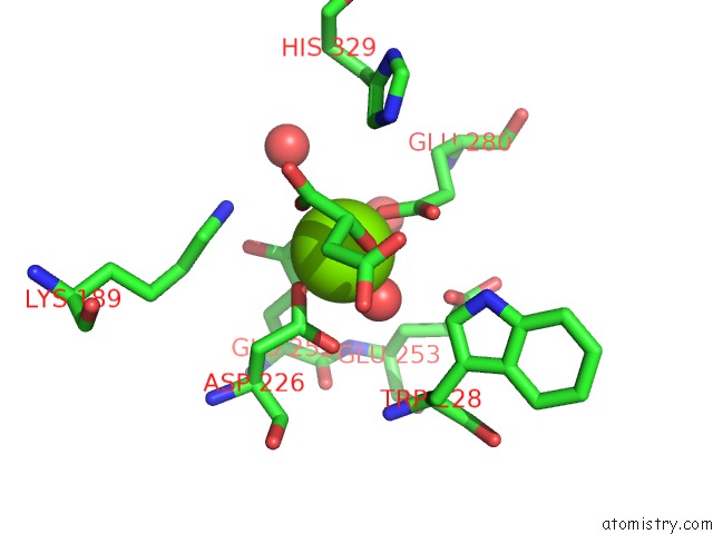
 Mono view
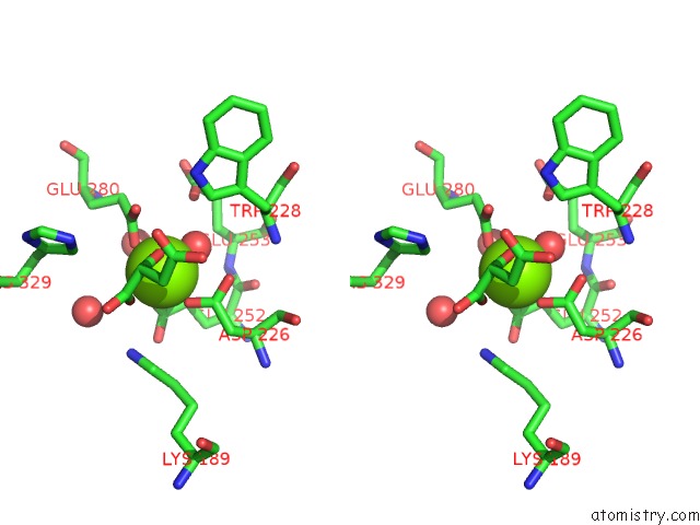
 Stereo pair view
|
A full contact list of Magnesium with other atoms in the Mg binding
site number 6 of Crystal Structure of L-Rhamnonate Dehydratase From Salmonella Typhimurium Complexed with Mg and D-Malate within 5.0Å range:
probe
|
atom
|
residue
|
distance (Å)
|
B
|
Occ
|
F:Mg501
b:14.2
occ:1.00
|
OE1
|
F:GLU280
|
2.0
|
15.1
|
1.0
|
OD2
|
F:ASP226
|
2.0
|
11.6
|
1.0
|
O
|
F:HOH542
|
2.0
|
13.4
|
1.0
|
OE1
|
F:GLU252
|
2.1
|
13.5
|
1.0
|
O3
|
F:MLT502
|
2.2
|
18.8
|
1.0
|
O1
|
F:MLT502
|
2.3
|
19.9
|
1.0
|
CD
|
F:GLU280
|
2.9
|
16.6
|
1.0
|
CG
|
F:ASP226
|
3.0
|
13.6
|
1.0
|
C1
|
F:MLT502
|
3.1
|
23.5
|
1.0
|
CD
|
F:GLU252
|
3.1
|
15.2
|
1.0
|
C2
|
F:MLT502
|
3.1
|
21.5
|
1.0
|
OE2
|
F:GLU280
|
3.3
|
15.6
|
1.0
|
OD1
|
F:ASP226
|
3.3
|
14.0
|
1.0
|
CG
|
F:GLU252
|
3.7
|
11.8
|
1.0
|
OE2
|
F:GLU252
|
4.0
|
12.6
|
1.0
|
C3
|
F:MLT502
|
4.1
|
24.1
|
1.0
|
NZ
|
F:LYS189
|
4.1
|
12.7
|
1.0
|
O
|
F:HOH520
|
4.1
|
14.2
|
1.0
|
OE1
|
F:GLU253
|
4.2
|
13.6
|
1.0
|
O
|
F:HOH536
|
4.2
|
17.3
|
1.0
|
CG
|
F:GLU280
|
4.3
|
16.3
|
1.0
|
O2
|
F:MLT502
|
4.3
|
20.7
|
1.0
|
CB
|
F:ASP226
|
4.3
|
12.2
|
1.0
|
CD2
|
F:HIS329
|
4.5
|
23.2
|
1.0
|
CE
|
F:LYS189
|
4.6
|
14.9
|
1.0
|
CB
|
F:GLU253
|
4.9
|
14.0
|
1.0
|
CD1
|
F:TRP228
|
4.9
|
14.6
|
1.0
|
CB
|
F:GLU252
|
5.0
|
11.5
|
1.0
|
|
Magnesium binding site 7 out
of 8 in 3d47
Go back to
Magnesium Binding Sites List in 3d47
Magnesium binding site 7 out
of 8 in the Crystal Structure of L-Rhamnonate Dehydratase From Salmonella Typhimurium Complexed with Mg and D-Malate
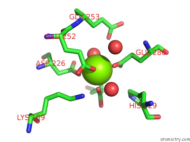
 Mono view
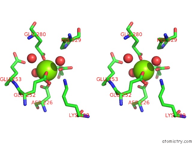
 Stereo pair view
|
A full contact list of Magnesium with other atoms in the Mg binding
site number 7 of Crystal Structure of L-Rhamnonate Dehydratase From Salmonella Typhimurium Complexed with Mg and D-Malate within 5.0Å range:
probe
|
atom
|
residue
|
distance (Å)
|
B
|
Occ
|
G:Mg501
b:15.9
occ:1.00
|
OE1
|
G:GLU280
|
2.0
|
14.5
|
1.0
|
O
|
G:HOH728
|
2.0
|
13.3
|
1.0
|
OE1
|
G:GLU252
|
2.0
|
12.0
|
1.0
|
OD2
|
G:ASP226
|
2.1
|
14.5
|
1.0
|
O1
|
G:MLT502
|
2.1
|
16.1
|
1.0
|
O3
|
G:MLT502
|
2.3
|
20.1
|
1.0
|
CD
|
G:GLU280
|
2.9
|
18.4
|
1.0
|
C1
|
G:MLT502
|
3.0
|
21.2
|
1.0
|
CD
|
G:GLU252
|
3.0
|
15.2
|
1.0
|
CG
|
G:ASP226
|
3.1
|
13.7
|
1.0
|
C2
|
G:MLT502
|
3.2
|
21.4
|
1.0
|
OE2
|
G:GLU280
|
3.3
|
18.0
|
1.0
|
OD1
|
G:ASP226
|
3.5
|
13.9
|
1.0
|
CG
|
G:GLU252
|
3.6
|
12.3
|
1.0
|
NZ
|
G:LYS189
|
3.9
|
12.6
|
1.0
|
O
|
G:HOH798
|
3.9
|
15.3
|
1.0
|
O
|
G:HOH799
|
4.0
|
17.0
|
1.0
|
OE2
|
G:GLU252
|
4.0
|
12.0
|
1.0
|
O2
|
G:MLT502
|
4.1
|
18.2
|
1.0
|
C3
|
G:MLT502
|
4.2
|
25.3
|
1.0
|
OE1
|
G:GLU253
|
4.2
|
16.9
|
1.0
|
CG
|
G:GLU280
|
4.2
|
16.0
|
1.0
|
CD2
|
G:HIS329
|
4.4
|
28.2
|
1.0
|
CB
|
G:ASP226
|
4.5
|
13.4
|
1.0
|
CE
|
G:LYS189
|
4.5
|
13.8
|
1.0
|
CB
|
G:GLU253
|
4.9
|
14.1
|
1.0
|
CB
|
G:GLU252
|
4.9
|
10.8
|
1.0
|
CB
|
G:GLU280
|
4.9
|
15.4
|
1.0
|
|
Magnesium binding site 8 out
of 8 in 3d47
Go back to
Magnesium Binding Sites List in 3d47
Magnesium binding site 8 out
of 8 in the Crystal Structure of L-Rhamnonate Dehydratase From Salmonella Typhimurium Complexed with Mg and D-Malate
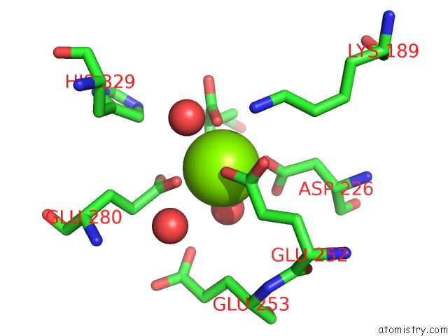
 Mono view
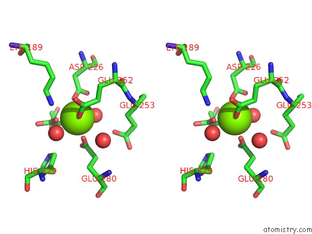
 Stereo pair view
|
A full contact list of Magnesium with other atoms in the Mg binding
site number 8 of Crystal Structure of L-Rhamnonate Dehydratase From Salmonella Typhimurium Complexed with Mg and D-Malate within 5.0Å range:
probe
|
atom
|
residue
|
distance (Å)
|
B
|
Occ
|
H:Mg501
b:22.9
occ:1.00
|
OE1
|
H:GLU252
|
1.9
|
16.3
|
1.0
|
O
|
H:HOH505
|
2.0
|
19.6
|
1.0
|
OD2
|
H:ASP226
|
2.1
|
20.1
|
1.0
|
O1
|
H:MLT502
|
2.1
|
28.4
|
1.0
|
OE1
|
H:GLU280
|
2.1
|
18.7
|
1.0
|
O3
|
H:MLT502
|
2.3
|
26.6
|
1.0
|
CD
|
H:GLU252
|
2.9
|
18.3
|
1.0
|
C1
|
H:MLT502
|
2.9
|
29.2
|
1.0
|
CD
|
H:GLU280
|
3.1
|
20.0
|
1.0
|
CG
|
H:ASP226
|
3.1
|
22.2
|
1.0
|
C2
|
H:MLT502
|
3.1
|
28.9
|
1.0
|
OE2
|
H:GLU280
|
3.4
|
19.9
|
1.0
|
OD1
|
H:ASP226
|
3.5
|
20.2
|
1.0
|
CG
|
H:GLU252
|
3.6
|
17.9
|
1.0
|
NZ
|
H:LYS189
|
3.7
|
17.3
|
1.0
|
OE2
|
H:GLU252
|
3.9
|
18.6
|
1.0
|
O
|
H:HOH522
|
4.0
|
20.3
|
1.0
|
O
|
H:HOH516
|
4.1
|
20.5
|
1.0
|
O2
|
H:MLT502
|
4.1
|
29.6
|
1.0
|
C3
|
H:MLT502
|
4.2
|
27.1
|
1.0
|
CD2
|
H:HIS329
|
4.3
|
26.6
|
1.0
|
CG
|
H:GLU280
|
4.4
|
19.7
|
1.0
|
OE1
|
H:GLU253
|
4.4
|
21.4
|
1.0
|
CB
|
H:ASP226
|
4.4
|
22.1
|
1.0
|
CE
|
H:LYS189
|
4.5
|
18.6
|
1.0
|
CB
|
H:GLU252
|
4.8
|
17.8
|
1.0
|
NE2
|
H:HIS329
|
4.9
|
28.1
|
1.0
|
|
Reference:
A.A.Fedorov,
E.V.Fedorov,
J.M.Sauder,
S.K.Burley,
J.A.Gerlt,
S.C.Almo.
Crystal Structure of L-Rhamnonate Dehydratase From Salmonella Typhimurium Complexed with Mg and D-Malate. To Be Published.
Page generated: Wed Aug 14 12:23:17 2024
|