Magnesium »
PDB 3d1r-3dev »
3d6t »
Magnesium in PDB 3d6t: Structure of the Roc Domain From the Parkinson'S Disease-Associated Leucine-Rich Repeat Kinase 2 Reveals A Dimeric Gtpase
Enzymatic activity of Structure of the Roc Domain From the Parkinson'S Disease-Associated Leucine-Rich Repeat Kinase 2 Reveals A Dimeric Gtpase
All present enzymatic activity of Structure of the Roc Domain From the Parkinson'S Disease-Associated Leucine-Rich Repeat Kinase 2 Reveals A Dimeric Gtpase:
2.7.11.1;
2.7.11.1;
Protein crystallography data
The structure of Structure of the Roc Domain From the Parkinson'S Disease-Associated Leucine-Rich Repeat Kinase 2 Reveals A Dimeric Gtpase, PDB code: 3d6t
was solved by
J.Deng,
with X-Ray Crystallography technique. A brief refinement statistics is given in the table below:
| Resolution Low / High (Å) | 21.09 / 2.43 |
| Space group | C 1 2 1 |
| Cell size a, b, c (Å), α, β, γ (°) | 69.919, 53.167, 42.736, 90.00, 112.76, 90.00 |
| R / Rfree (%) | 21.1 / 30.7 |
Magnesium Binding Sites:
The binding sites of Magnesium atom in the Structure of the Roc Domain From the Parkinson'S Disease-Associated Leucine-Rich Repeat Kinase 2 Reveals A Dimeric Gtpase
(pdb code 3d6t). This binding sites where shown within
5.0 Angstroms radius around Magnesium atom.
In total only one binding site of Magnesium was determined in the Structure of the Roc Domain From the Parkinson'S Disease-Associated Leucine-Rich Repeat Kinase 2 Reveals A Dimeric Gtpase, PDB code: 3d6t:
In total only one binding site of Magnesium was determined in the Structure of the Roc Domain From the Parkinson'S Disease-Associated Leucine-Rich Repeat Kinase 2 Reveals A Dimeric Gtpase, PDB code: 3d6t:
Magnesium binding site 1 out of 1 in 3d6t
Go back to
Magnesium binding site 1 out
of 1 in the Structure of the Roc Domain From the Parkinson'S Disease-Associated Leucine-Rich Repeat Kinase 2 Reveals A Dimeric Gtpase
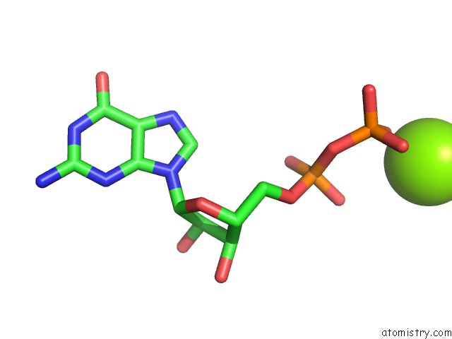
Mono view
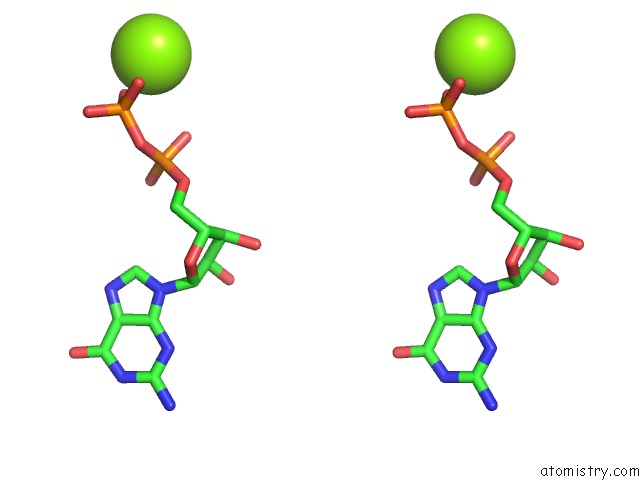
Stereo pair view
Mono view
Stereo pair view
A full contact list of Magnesium with other atoms in the Mg binding
site number 1 of Structure of the Roc Domain From the Parkinson'S Disease-Associated Leucine-Rich Repeat Kinase 2 Reveals A Dimeric Gtpase within 5.0Å range:
|
Reference:
J.Deng,
P.A.Lewis,
E.Greggio,
E.Sluch,
A.Beilina,
M.R.Cookson.
Structure of the Roc Domain From the Parkinson'S Disease-Associated Leucine-Rich Repeat Kinase 2 Reveals A Dimeric Gtpase Proc.Natl.Acad.Sci.Usa V. 105 1499 2008.
ISSN: ISSN 0027-8424
PubMed: 18230735
DOI: 10.1073/PNAS.0709098105
Page generated: Wed Aug 14 12:24:11 2024
ISSN: ISSN 0027-8424
PubMed: 18230735
DOI: 10.1073/PNAS.0709098105
Last articles
Zn in 9J0NZn in 9J0O
Zn in 9J0P
Zn in 9FJX
Zn in 9EKB
Zn in 9C0F
Zn in 9CAH
Zn in 9CH0
Zn in 9CH3
Zn in 9CH1