Magnesium »
PDB 3d1r-3dev »
3da3 »
Magnesium in PDB 3da3: Crystal Structure of Colicin M, A Novel Phosphatase Specifically Imported By Escherichia Coli
Protein crystallography data
The structure of Crystal Structure of Colicin M, A Novel Phosphatase Specifically Imported By Escherichia Coli, PDB code: 3da3
was solved by
K.Zeth,
R.Albrecht,
C.Romer,
V.Braun,
with X-Ray Crystallography technique. A brief refinement statistics is given in the table below:
| Resolution Low / High (Å) | 19.98 / 2.50 |
| Space group | P 42 21 2 |
| Cell size a, b, c (Å), α, β, γ (°) | 119.880, 119.880, 96.220, 90.00, 90.00, 90.00 |
| R / Rfree (%) | 23.5 / 29.5 |
Magnesium Binding Sites:
The binding sites of Magnesium atom in the Crystal Structure of Colicin M, A Novel Phosphatase Specifically Imported By Escherichia Coli
(pdb code 3da3). This binding sites where shown within
5.0 Angstroms radius around Magnesium atom.
In total 2 binding sites of Magnesium where determined in the Crystal Structure of Colicin M, A Novel Phosphatase Specifically Imported By Escherichia Coli, PDB code: 3da3:
Jump to Magnesium binding site number: 1; 2;
In total 2 binding sites of Magnesium where determined in the Crystal Structure of Colicin M, A Novel Phosphatase Specifically Imported By Escherichia Coli, PDB code: 3da3:
Jump to Magnesium binding site number: 1; 2;
Magnesium binding site 1 out of 2 in 3da3
Go back to
Magnesium binding site 1 out
of 2 in the Crystal Structure of Colicin M, A Novel Phosphatase Specifically Imported By Escherichia Coli
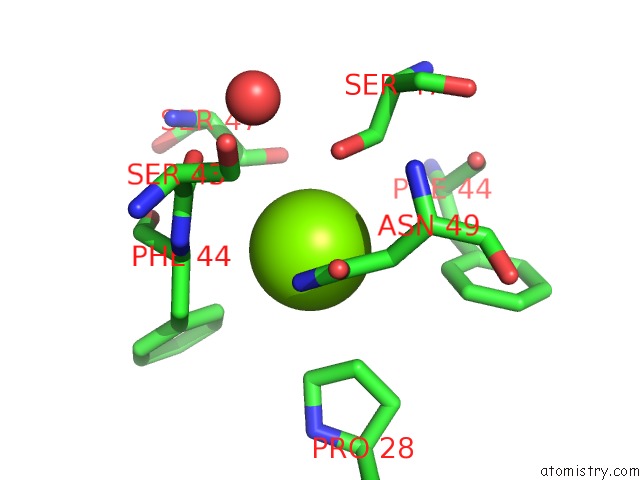
Mono view
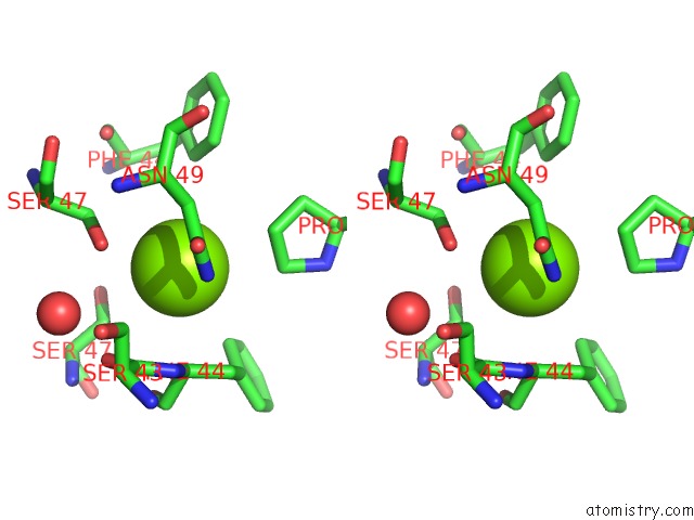
Stereo pair view
Mono view
Stereo pair view
A full contact list of Magnesium with other atoms in the Mg binding
site number 1 of Crystal Structure of Colicin M, A Novel Phosphatase Specifically Imported By Escherichia Coli within 5.0Å range:
|
Magnesium binding site 2 out of 2 in 3da3
Go back to
Magnesium binding site 2 out
of 2 in the Crystal Structure of Colicin M, A Novel Phosphatase Specifically Imported By Escherichia Coli
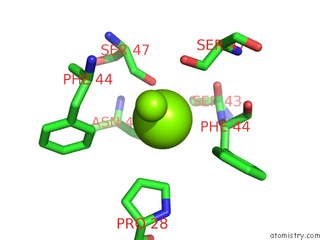
Mono view
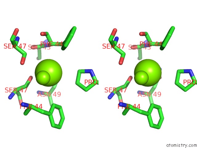
Stereo pair view
Mono view
Stereo pair view
A full contact list of Magnesium with other atoms in the Mg binding
site number 2 of Crystal Structure of Colicin M, A Novel Phosphatase Specifically Imported By Escherichia Coli within 5.0Å range:
|
Reference:
K.Zeth,
C.Romer,
S.I.Patzer,
V.Braun.
Crystal Structure of Colicin M, A Novel Phosphatase Specifically Imported By Escherichia Coli J.Biol.Chem. V. 283 25324 2008.
ISSN: ISSN 0021-9258
PubMed: 18640984
DOI: 10.1074/JBC.M802591200
Page generated: Wed Aug 14 12:25:08 2024
ISSN: ISSN 0021-9258
PubMed: 18640984
DOI: 10.1074/JBC.M802591200
Last articles
Zn in 9J0NZn in 9J0O
Zn in 9J0P
Zn in 9FJX
Zn in 9EKB
Zn in 9C0F
Zn in 9CAH
Zn in 9CH0
Zn in 9CH3
Zn in 9CH1