Magnesium »
PDB 3dfy-3dts »
3dgt »
Magnesium in PDB 3dgt: The 1.5 A Crystal Structure of Endo-1,3-Beta-Glucanase From Streptomyces Sioyaensis
Enzymatic activity of The 1.5 A Crystal Structure of Endo-1,3-Beta-Glucanase From Streptomyces Sioyaensis
All present enzymatic activity of The 1.5 A Crystal Structure of Endo-1,3-Beta-Glucanase From Streptomyces Sioyaensis:
3.2.1.39;
3.2.1.39;
Protein crystallography data
The structure of The 1.5 A Crystal Structure of Endo-1,3-Beta-Glucanase From Streptomyces Sioyaensis, PDB code: 3dgt
was solved by
T.H.Li,
with X-Ray Crystallography technique. A brief refinement statistics is given in the table below:
| Resolution Low / High (Å) | 19.75 / 1.50 |
| Space group | P 21 21 21 |
| Cell size a, b, c (Å), α, β, γ (°) | 39.509, 75.967, 79.663, 90.00, 90.00, 90.00 |
| R / Rfree (%) | 18.3 / 19.8 |
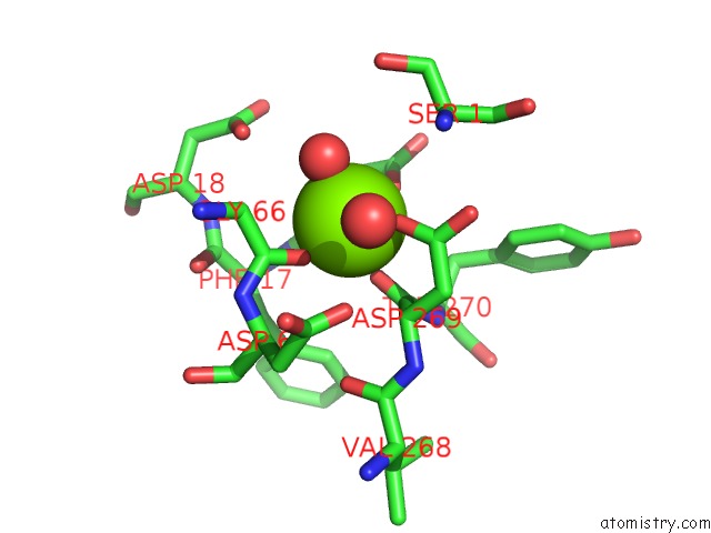
Magnesium Binding Sites:
The binding sites of Magnesium atom in the The 1.5 A Crystal Structure of Endo-1,3-Beta-Glucanase From Streptomyces Sioyaensis
(pdb code 3dgt). This binding sites where shown within
5.0 Angstroms radius around Magnesium atom.
In total only one binding site of Magnesium was determined in the The 1.5 A Crystal Structure of Endo-1,3-Beta-Glucanase From Streptomyces Sioyaensis, PDB code: 3dgt:
In total only one binding site of Magnesium was determined in the The 1.5 A Crystal Structure of Endo-1,3-Beta-Glucanase From Streptomyces Sioyaensis, PDB code: 3dgt:
Magnesium binding site 1 out of 1 in 3dgt
Go back to
Magnesium binding site 1 out
of 1 in the The 1.5 A Crystal Structure of Endo-1,3-Beta-Glucanase From Streptomyces Sioyaensis
Mono view
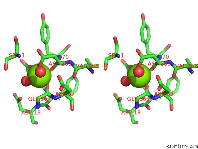
Stereo pair view
Mono view
Stereo pair view
A full contact list of Magnesium with other atoms in the Mg binding
site number 1 of The 1.5 A Crystal Structure of Endo-1,3-Beta-Glucanase From Streptomyces Sioyaensis within 5.0Å range:
|
Reference:
T.-Y.Hong,
Y.-Y.Hsiao,
M.Meng,
T.T.Li.
The 1.5 A Structure of Endo-1,3-Beta-Glucanase From Streptomyces Sioyaensis: Evolution of the Active-Site Structure For 1,3-Beta-Glucan-Binding Specificity and Hydrolysis Acta Crystallogr.,Sect.D V. 64 964 2008.
ISSN: ISSN 0907-4449
PubMed: 18703845
DOI: 10.1107/S0907444908021550
Page generated: Wed Aug 14 12:30:32 2024
ISSN: ISSN 0907-4449
PubMed: 18703845
DOI: 10.1107/S0907444908021550
Last articles
Zn in 9J0NZn in 9J0O
Zn in 9J0P
Zn in 9FJX
Zn in 9EKB
Zn in 9C0F
Zn in 9CAH
Zn in 9CH0
Zn in 9CH3
Zn in 9CH1