Magnesium »
PDB 3dfy-3dts »
3dil »
Magnesium in PDB 3dil: Crystal Structure of the Thermotoga Maritima Lysine Riboswitch Bound to Lysine
Protein crystallography data
The structure of Crystal Structure of the Thermotoga Maritima Lysine Riboswitch Bound to Lysine, PDB code: 3dil
was solved by
A.A.Serganov,
with X-Ray Crystallography technique. A brief refinement statistics is given in the table below:
| Resolution Low / High (Å) | 20.00 / 1.90 |
| Space group | P 21 21 21 |
| Cell size a, b, c (Å), α, β, γ (°) | 54.216, 78.953, 139.988, 90.00, 90.00, 90.00 |
| R / Rfree (%) | 19.2 / 22.9 |
Other elements in 3dil:
The structure of Crystal Structure of the Thermotoga Maritima Lysine Riboswitch Bound to Lysine also contains other interesting chemical elements:
| Potassium | (K) | 3 atoms |
| Sodium | (Na) | 29 atoms |
Magnesium Binding Sites:
The binding sites of Magnesium atom in the Crystal Structure of the Thermotoga Maritima Lysine Riboswitch Bound to Lysine
(pdb code 3dil). This binding sites where shown within
5.0 Angstroms radius around Magnesium atom.
In total only one binding site of Magnesium was determined in the Crystal Structure of the Thermotoga Maritima Lysine Riboswitch Bound to Lysine, PDB code: 3dil:
In total only one binding site of Magnesium was determined in the Crystal Structure of the Thermotoga Maritima Lysine Riboswitch Bound to Lysine, PDB code: 3dil:
Magnesium binding site 1 out of 1 in 3dil
Go back to
Magnesium binding site 1 out
of 1 in the Crystal Structure of the Thermotoga Maritima Lysine Riboswitch Bound to Lysine
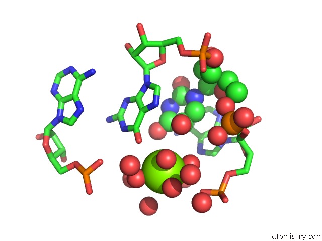
Mono view
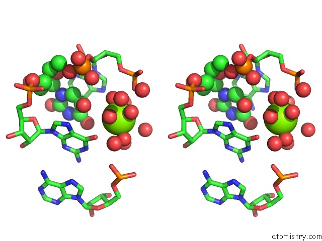
Stereo pair view
Mono view
Stereo pair view
A full contact list of Magnesium with other atoms in the Mg binding
site number 1 of Crystal Structure of the Thermotoga Maritima Lysine Riboswitch Bound to Lysine within 5.0Å range:
|
Reference:
A.Serganov,
L.Huang,
D.J.Patel.
Structural Insights Into Amino Acid Binding and Gene Control By A Lysine Riboswitch. Nature V. 455 1263 2008.
ISSN: ISSN 0028-0836
PubMed: 18784651
DOI: 10.1038/NATURE07326
Page generated: Wed Aug 14 12:31:00 2024
ISSN: ISSN 0028-0836
PubMed: 18784651
DOI: 10.1038/NATURE07326
Last articles
Zn in 9J0NZn in 9J0O
Zn in 9J0P
Zn in 9FJX
Zn in 9EKB
Zn in 9C0F
Zn in 9CAH
Zn in 9CH0
Zn in 9CH3
Zn in 9CH1