Magnesium »
PDB 3dfy-3dts »
3dmi »
Magnesium in PDB 3dmi: Crystallization and Structural Analysis of Cytochrome C6 From the Diatom Phaeodactylum Tricornutum at 1.5 A Resolution
Protein crystallography data
The structure of Crystallization and Structural Analysis of Cytochrome C6 From the Diatom Phaeodactylum Tricornutum at 1.5 A Resolution, PDB code: 3dmi
was solved by
H.Akazaki,
F.Kawai,
M.Hosokawa,
T.Hama,
T.Hirano,
B.-K.Lim,
N.Sakurai,
W.Hakamata,
S.-Y.Park,
T.Nishio,
T.Oku,
with X-Ray Crystallography technique. A brief refinement statistics is given in the table below:
| Resolution Low / High (Å) | 20.00 / 1.50 |
| Space group | I 2 3 |
| Cell size a, b, c (Å), α, β, γ (°) | 80.385, 80.385, 80.385, 90.00, 90.00, 90.00 |
| R / Rfree (%) | 16.4 / 20.2 |
Other elements in 3dmi:
The structure of Crystallization and Structural Analysis of Cytochrome C6 From the Diatom Phaeodactylum Tricornutum at 1.5 A Resolution also contains other interesting chemical elements:
| Iron | (Fe) | 1 atom |
Magnesium Binding Sites:
The binding sites of Magnesium atom in the Crystallization and Structural Analysis of Cytochrome C6 From the Diatom Phaeodactylum Tricornutum at 1.5 A Resolution
(pdb code 3dmi). This binding sites where shown within
5.0 Angstroms radius around Magnesium atom.
In total only one binding site of Magnesium was determined in the Crystallization and Structural Analysis of Cytochrome C6 From the Diatom Phaeodactylum Tricornutum at 1.5 A Resolution, PDB code: 3dmi:
In total only one binding site of Magnesium was determined in the Crystallization and Structural Analysis of Cytochrome C6 From the Diatom Phaeodactylum Tricornutum at 1.5 A Resolution, PDB code: 3dmi:
Magnesium binding site 1 out of 1 in 3dmi
Go back to
Magnesium binding site 1 out
of 1 in the Crystallization and Structural Analysis of Cytochrome C6 From the Diatom Phaeodactylum Tricornutum at 1.5 A Resolution
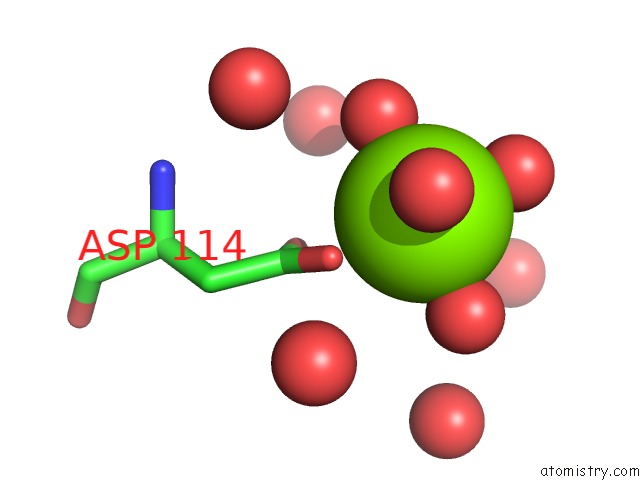
Mono view
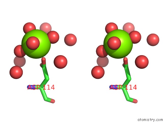
Stereo pair view
Mono view
Stereo pair view
A full contact list of Magnesium with other atoms in the Mg binding
site number 1 of Crystallization and Structural Analysis of Cytochrome C6 From the Diatom Phaeodactylum Tricornutum at 1.5 A Resolution within 5.0Å range:
|
Reference:
H.Akazaki,
F.Kawai,
M.Hosokawa,
T.Hama,
H.Chida,
T.Hirano,
B.K.Lim,
N.Sakurai,
W.Hakamata,
S.Y.Park,
T.Nishio,
T.Oku.
Crystallization and Structural Analysis of Cytochrome C(6) From the Diatom Phaeodactylum Tricornutum at 1.5 A Resolution. Biosci.Biotechnol.Biochem. V. 73 189 2009.
ISSN: ISSN 0916-8451
PubMed: 19129656
DOI: 10.1271/BBB.80472
Page generated: Wed Aug 14 12:34:26 2024
ISSN: ISSN 0916-8451
PubMed: 19129656
DOI: 10.1271/BBB.80472
Last articles
Zn in 9J0NZn in 9J0O
Zn in 9J0P
Zn in 9FJX
Zn in 9EKB
Zn in 9C0F
Zn in 9CAH
Zn in 9CH0
Zn in 9CH3
Zn in 9CH1