Magnesium »
PDB 3dtu-3e35 »
3duq »
Magnesium in PDB 3duq: E(L212)A, D(L213)A, N(M5)D Triple Mutant Structure of Photosynthetic Reaction Center From Rhodobacter Sphaeroides
Protein crystallography data
The structure of E(L212)A, D(L213)A, N(M5)D Triple Mutant Structure of Photosynthetic Reaction Center From Rhodobacter Sphaeroides, PDB code: 3duq
was solved by
P.R.Pokkuluri,
M.Schiffer,
with X-Ray Crystallography technique. A brief refinement statistics is given in the table below:
| Resolution Low / High (Å) | 30.00 / 2.70 |
| Space group | P 31 2 1 |
| Cell size a, b, c (Å), α, β, γ (°) | 141.300, 141.300, 187.400, 90.00, 90.00, 120.00 |
| R / Rfree (%) | 20.9 / 21.7 |
Other elements in 3duq:
The structure of E(L212)A, D(L213)A, N(M5)D Triple Mutant Structure of Photosynthetic Reaction Center From Rhodobacter Sphaeroides also contains other interesting chemical elements:
| Iron | (Fe) | 1 atom |
Magnesium Binding Sites:
The binding sites of Magnesium atom in the E(L212)A, D(L213)A, N(M5)D Triple Mutant Structure of Photosynthetic Reaction Center From Rhodobacter Sphaeroides
(pdb code 3duq). This binding sites where shown within
5.0 Angstroms radius around Magnesium atom.
In total 4 binding sites of Magnesium where determined in the E(L212)A, D(L213)A, N(M5)D Triple Mutant Structure of Photosynthetic Reaction Center From Rhodobacter Sphaeroides, PDB code: 3duq:
Jump to Magnesium binding site number: 1; 2; 3; 4;
In total 4 binding sites of Magnesium where determined in the E(L212)A, D(L213)A, N(M5)D Triple Mutant Structure of Photosynthetic Reaction Center From Rhodobacter Sphaeroides, PDB code: 3duq:
Jump to Magnesium binding site number: 1; 2; 3; 4;
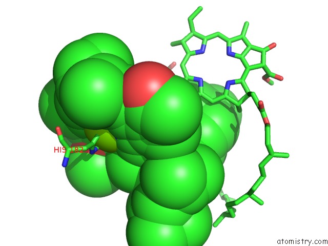
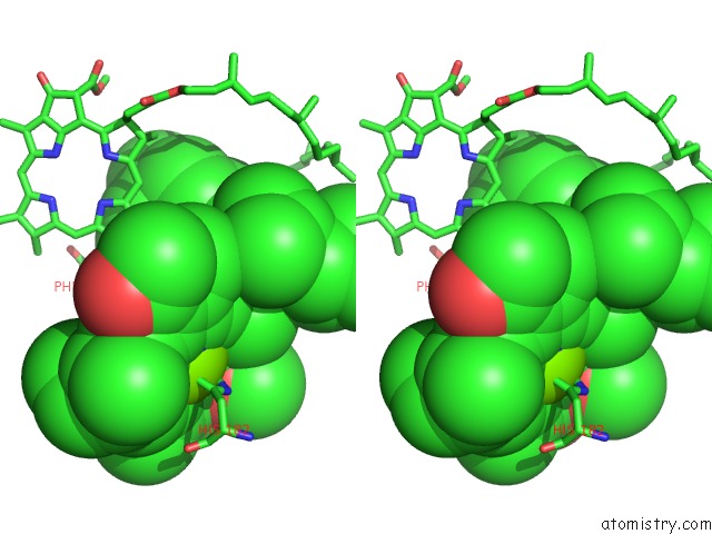
Magnesium binding site 1 out of 4 in 3duq
Go back to
Magnesium binding site 1 out
of 4 in the E(L212)A, D(L213)A, N(M5)D Triple Mutant Structure of Photosynthetic Reaction Center From Rhodobacter Sphaeroides
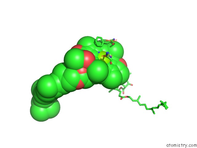
Mono view
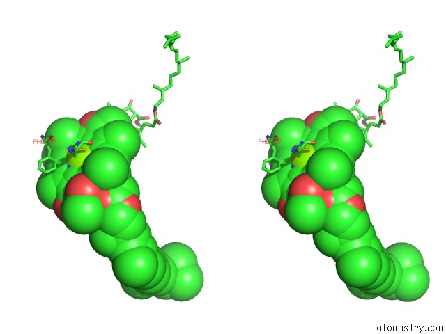
Stereo pair view
Mono view
Stereo pair view
A full contact list of Magnesium with other atoms in the Mg binding
site number 1 of E(L212)A, D(L213)A, N(M5)D Triple Mutant Structure of Photosynthetic Reaction Center From Rhodobacter Sphaeroides within 5.0Å range:
|
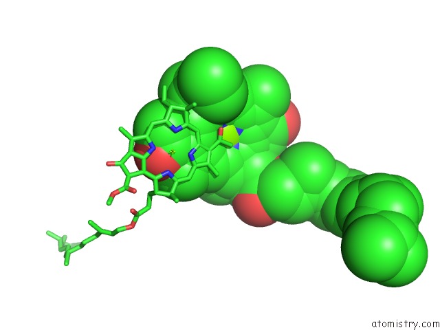
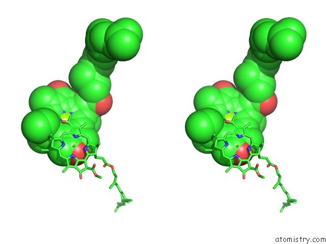
Magnesium binding site 2 out of 4 in 3duq
Go back to
Magnesium binding site 2 out
of 4 in the E(L212)A, D(L213)A, N(M5)D Triple Mutant Structure of Photosynthetic Reaction Center From Rhodobacter Sphaeroides
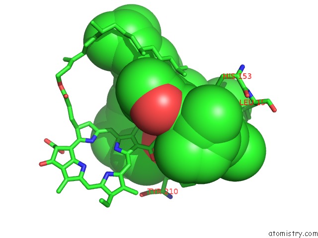
Mono view
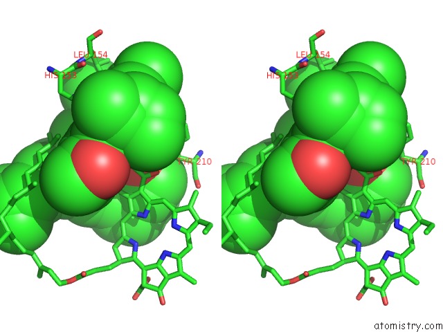
Stereo pair view
Mono view
Stereo pair view
A full contact list of Magnesium with other atoms in the Mg binding
site number 2 of E(L212)A, D(L213)A, N(M5)D Triple Mutant Structure of Photosynthetic Reaction Center From Rhodobacter Sphaeroides within 5.0Å range:
|
Magnesium binding site 3 out of 4 in 3duq
Go back to
Magnesium binding site 3 out
of 4 in the E(L212)A, D(L213)A, N(M5)D Triple Mutant Structure of Photosynthetic Reaction Center From Rhodobacter Sphaeroides
Mono view
Stereo pair view
Mono view
Stereo pair view
A full contact list of Magnesium with other atoms in the Mg binding
site number 3 of E(L212)A, D(L213)A, N(M5)D Triple Mutant Structure of Photosynthetic Reaction Center From Rhodobacter Sphaeroides within 5.0Å range:
|
Magnesium binding site 4 out of 4 in 3duq
Go back to
Magnesium binding site 4 out
of 4 in the E(L212)A, D(L213)A, N(M5)D Triple Mutant Structure of Photosynthetic Reaction Center From Rhodobacter Sphaeroides
Mono view
Stereo pair view
Mono view
Stereo pair view
A full contact list of Magnesium with other atoms in the Mg binding
site number 4 of E(L212)A, D(L213)A, N(M5)D Triple Mutant Structure of Photosynthetic Reaction Center From Rhodobacter Sphaeroides within 5.0Å range:
|
Reference:
P.R.Pokkuluri,
P.D.Laible,
S.L.Ginell,
D.K.Hanson,
M.Schiffer.
Structural Description of Compensatory Mutations That Restore Proton Transfer Pathways to the L212A-L213A Mutant Bacterial Reaction Center To Be Published.
Page generated: Wed Aug 14 12:42:52 2024
Last articles
Zn in 9JYWZn in 9IR4
Zn in 9IR3
Zn in 9GMX
Zn in 9GMW
Zn in 9JEJ
Zn in 9ERF
Zn in 9ERE
Zn in 9EGV
Zn in 9EGW