Magnesium »
PDB 3e3z-3ef8 »
3edn »
Magnesium in PDB 3edn: Crystal Structure of the Bacillus Anthracis Phenazine Biosynthesis Protein, Phzf Family
Protein crystallography data
The structure of Crystal Structure of the Bacillus Anthracis Phenazine Biosynthesis Protein, Phzf Family, PDB code: 3edn
was solved by
S.M.Anderson,
J.S.Brunzelle,
O.Onopriyenko,
A.Savchenko,
W.F.Anderson,
Center For Structural Genomics Of Infectiousdiseases (Csgid),
with X-Ray Crystallography technique. A brief refinement statistics is given in the table below:
| Resolution Low / High (Å) | 38.18 / 1.50 |
| Space group | P 21 21 21 |
| Cell size a, b, c (Å), α, β, γ (°) | 60.089, 65.638, 150.170, 90.00, 90.00, 90.00 |
| R / Rfree (%) | 18 / 21.9 |
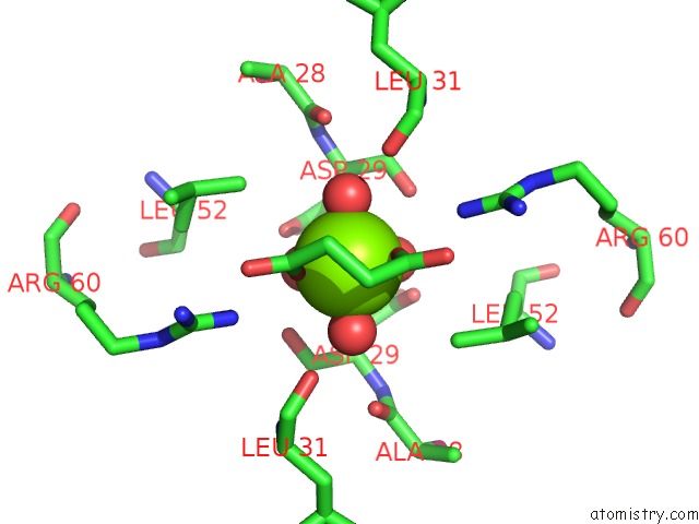
Magnesium Binding Sites:
The binding sites of Magnesium atom in the Crystal Structure of the Bacillus Anthracis Phenazine Biosynthesis Protein, Phzf Family
(pdb code 3edn). This binding sites where shown within
5.0 Angstroms radius around Magnesium atom.
In total only one binding site of Magnesium was determined in the Crystal Structure of the Bacillus Anthracis Phenazine Biosynthesis Protein, Phzf Family, PDB code: 3edn:
In total only one binding site of Magnesium was determined in the Crystal Structure of the Bacillus Anthracis Phenazine Biosynthesis Protein, Phzf Family, PDB code: 3edn:
Magnesium binding site 1 out of 1 in 3edn
Go back to
Magnesium binding site 1 out
of 1 in the Crystal Structure of the Bacillus Anthracis Phenazine Biosynthesis Protein, Phzf Family
Mono view

Stereo pair view
Mono view
Stereo pair view
A full contact list of Magnesium with other atoms in the Mg binding
site number 1 of Crystal Structure of the Bacillus Anthracis Phenazine Biosynthesis Protein, Phzf Family within 5.0Å range:
|
Reference:
S.M.Anderson,
J.S.Brunzelle,
O.Onopriyenko,
A.Savchenko,
W.F.Anderson.
Crystal Structure of the Bacillus Anthracis Phenazine Biosynthesis Protein, Phzf Family To Be Published.
Page generated: Sun Aug 10 20:27:49 2025
Last articles
Mg in 6K27Mg in 6K7K
Mg in 6K7J
Mg in 6K77
Mg in 6K74
Mg in 6K55
Mg in 6K5P
Mg in 6K4E
Mg in 6K4Y
Mg in 6K32