Magnesium »
PDB 3e3z-3ef8 »
3ee3 »
Magnesium in PDB 3ee3: Crystal Structure of Acanthamoeba Polyphaga Mimivirus Nucleoside Diphosphate Kinase Complexed with Cdp
Enzymatic activity of Crystal Structure of Acanthamoeba Polyphaga Mimivirus Nucleoside Diphosphate Kinase Complexed with Cdp
All present enzymatic activity of Crystal Structure of Acanthamoeba Polyphaga Mimivirus Nucleoside Diphosphate Kinase Complexed with Cdp:
2.7.4.6;
2.7.4.6;
Protein crystallography data
The structure of Crystal Structure of Acanthamoeba Polyphaga Mimivirus Nucleoside Diphosphate Kinase Complexed with Cdp, PDB code: 3ee3
was solved by
S.Jeudy,
A.Lartigue,
J.M.Claverie,
C.Abergel,
with X-Ray Crystallography technique. A brief refinement statistics is given in the table below:
| Resolution Low / High (Å) | 20.00 / 2.40 |
| Space group | C 2 2 21 |
| Cell size a, b, c (Å), α, β, γ (°) | 82.419, 154.419, 187.039, 90.00, 90.00, 90.00 |
| R / Rfree (%) | 19.4 / 23.3 |
Magnesium Binding Sites:
The binding sites of Magnesium atom in the Crystal Structure of Acanthamoeba Polyphaga Mimivirus Nucleoside Diphosphate Kinase Complexed with Cdp
(pdb code 3ee3). This binding sites where shown within
5.0 Angstroms radius around Magnesium atom.
In total 6 binding sites of Magnesium where determined in the Crystal Structure of Acanthamoeba Polyphaga Mimivirus Nucleoside Diphosphate Kinase Complexed with Cdp, PDB code: 3ee3:
Jump to Magnesium binding site number: 1; 2; 3; 4; 5; 6;
In total 6 binding sites of Magnesium where determined in the Crystal Structure of Acanthamoeba Polyphaga Mimivirus Nucleoside Diphosphate Kinase Complexed with Cdp, PDB code: 3ee3:
Jump to Magnesium binding site number: 1; 2; 3; 4; 5; 6;
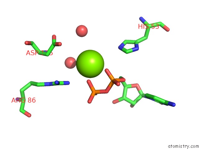
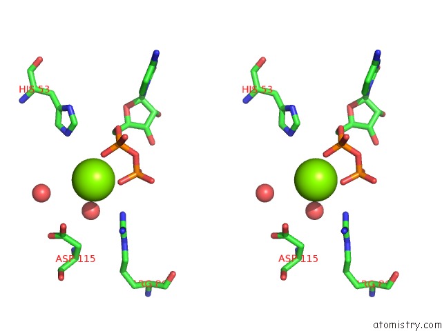
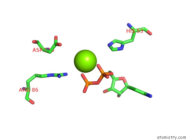
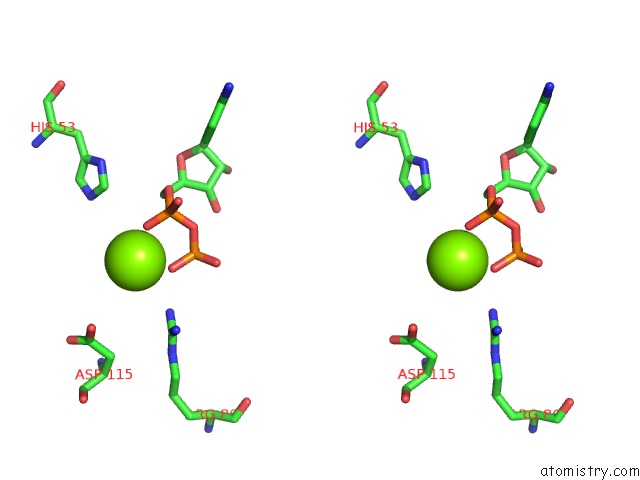
Magnesium binding site 1 out of 6 in 3ee3
Go back to
Magnesium binding site 1 out
of 6 in the Crystal Structure of Acanthamoeba Polyphaga Mimivirus Nucleoside Diphosphate Kinase Complexed with Cdp
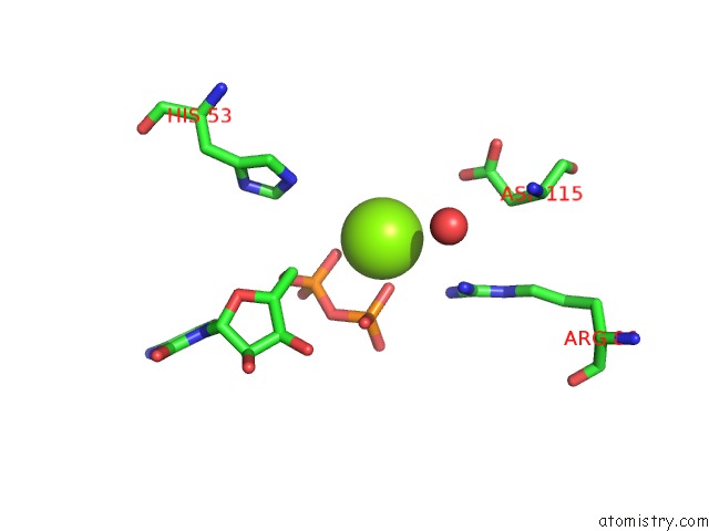
Mono view
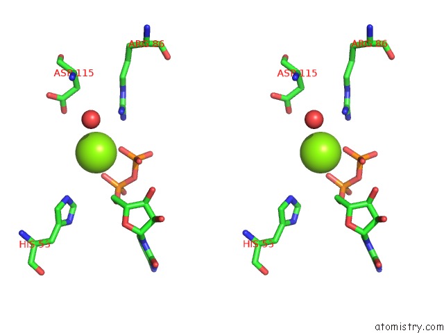
Stereo pair view
Mono view
Stereo pair view
A full contact list of Magnesium with other atoms in the Mg binding
site number 1 of Crystal Structure of Acanthamoeba Polyphaga Mimivirus Nucleoside Diphosphate Kinase Complexed with Cdp within 5.0Å range:
|
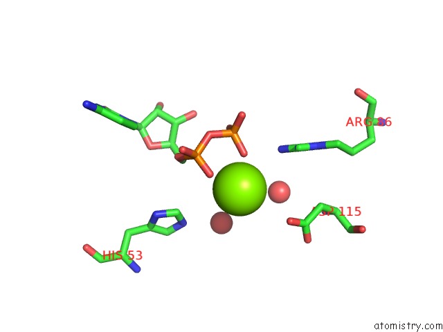
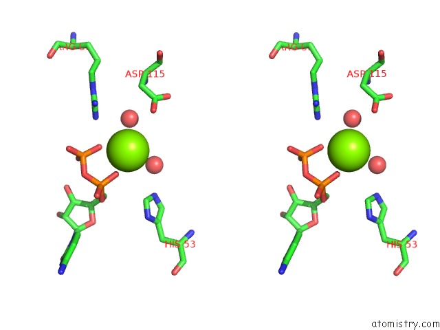
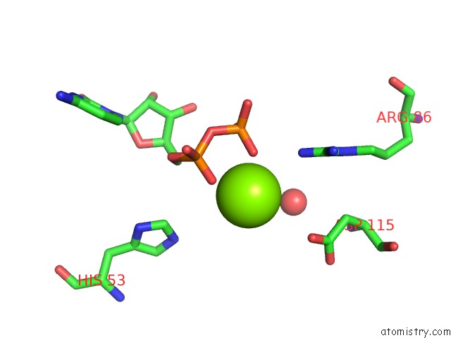
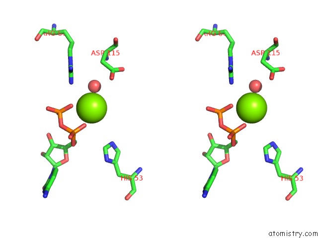
Magnesium binding site 2 out of 6 in 3ee3
Go back to
Magnesium binding site 2 out
of 6 in the Crystal Structure of Acanthamoeba Polyphaga Mimivirus Nucleoside Diphosphate Kinase Complexed with Cdp
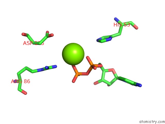
Mono view
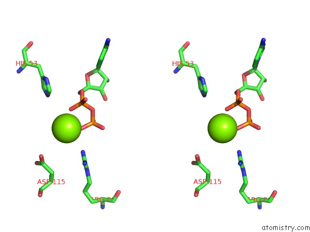
Stereo pair view
Mono view
Stereo pair view
A full contact list of Magnesium with other atoms in the Mg binding
site number 2 of Crystal Structure of Acanthamoeba Polyphaga Mimivirus Nucleoside Diphosphate Kinase Complexed with Cdp within 5.0Å range:
|
Magnesium binding site 3 out of 6 in 3ee3
Go back to
Magnesium binding site 3 out
of 6 in the Crystal Structure of Acanthamoeba Polyphaga Mimivirus Nucleoside Diphosphate Kinase Complexed with Cdp
Mono view
Stereo pair view
Mono view
Stereo pair view
A full contact list of Magnesium with other atoms in the Mg binding
site number 3 of Crystal Structure of Acanthamoeba Polyphaga Mimivirus Nucleoside Diphosphate Kinase Complexed with Cdp within 5.0Å range:
|
Magnesium binding site 4 out of 6 in 3ee3
Go back to
Magnesium binding site 4 out
of 6 in the Crystal Structure of Acanthamoeba Polyphaga Mimivirus Nucleoside Diphosphate Kinase Complexed with Cdp
Mono view
Stereo pair view
Mono view
Stereo pair view
A full contact list of Magnesium with other atoms in the Mg binding
site number 4 of Crystal Structure of Acanthamoeba Polyphaga Mimivirus Nucleoside Diphosphate Kinase Complexed with Cdp within 5.0Å range:
|
Magnesium binding site 5 out of 6 in 3ee3
Go back to
Magnesium binding site 5 out
of 6 in the Crystal Structure of Acanthamoeba Polyphaga Mimivirus Nucleoside Diphosphate Kinase Complexed with Cdp
Mono view
Stereo pair view
Mono view
Stereo pair view
A full contact list of Magnesium with other atoms in the Mg binding
site number 5 of Crystal Structure of Acanthamoeba Polyphaga Mimivirus Nucleoside Diphosphate Kinase Complexed with Cdp within 5.0Å range:
|
Magnesium binding site 6 out of 6 in 3ee3
Go back to
Magnesium binding site 6 out
of 6 in the Crystal Structure of Acanthamoeba Polyphaga Mimivirus Nucleoside Diphosphate Kinase Complexed with Cdp
Mono view
Stereo pair view
Mono view
Stereo pair view
A full contact list of Magnesium with other atoms in the Mg binding
site number 6 of Crystal Structure of Acanthamoeba Polyphaga Mimivirus Nucleoside Diphosphate Kinase Complexed with Cdp within 5.0Å range:
|
Reference:
S.Jeudy,
A.Lartigue,
J.M.Claverie,
C.Abergel.
Dissecting the Unique Nucleotide Specificity of Mimivirus Nucleoside Diphosphate Kinase. J.Virol. V. 83 7142 2009.
ISSN: ISSN 0022-538X
PubMed: 19439473
DOI: 10.1128/JVI.00511-09
Page generated: Sun Aug 10 20:28:17 2025
ISSN: ISSN 0022-538X
PubMed: 19439473
DOI: 10.1128/JVI.00511-09
Last articles
Mg in 6K27Mg in 6K7K
Mg in 6K7J
Mg in 6K77
Mg in 6K74
Mg in 6K55
Mg in 6K5P
Mg in 6K4E
Mg in 6K4Y
Mg in 6K32