Magnesium »
PDB 3efq-3eql »
3ehg »
Magnesium in PDB 3ehg: Crystal Structure of the Atp-Binding Domain of Desk in Complex with Atp
Enzymatic activity of Crystal Structure of the Atp-Binding Domain of Desk in Complex with Atp
All present enzymatic activity of Crystal Structure of the Atp-Binding Domain of Desk in Complex with Atp:
2.7.13.3;
2.7.13.3;
Protein crystallography data
The structure of Crystal Structure of the Atp-Binding Domain of Desk in Complex with Atp, PDB code: 3ehg
was solved by
F.Trajtenberg,
A.Buschiazzo,
with X-Ray Crystallography technique. A brief refinement statistics is given in the table below:
| Resolution Low / High (Å) | 42.03 / 1.74 |
| Space group | P 21 21 21 |
| Cell size a, b, c (Å), α, β, γ (°) | 40.379, 49.078, 81.436, 90.00, 90.00, 90.00 |
| R / Rfree (%) | 16.4 / 21.5 |
Other elements in 3ehg:
The structure of Crystal Structure of the Atp-Binding Domain of Desk in Complex with Atp also contains other interesting chemical elements:
| Iodine | (I) | 12 atoms |
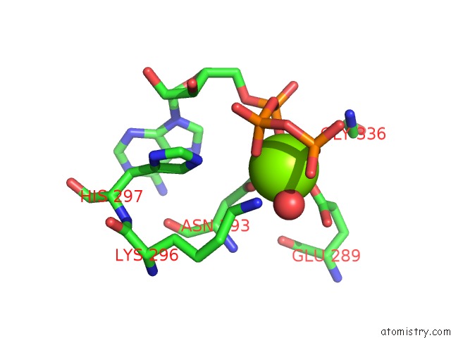
Magnesium Binding Sites:
The binding sites of Magnesium atom in the Crystal Structure of the Atp-Binding Domain of Desk in Complex with Atp
(pdb code 3ehg). This binding sites where shown within
5.0 Angstroms radius around Magnesium atom.
In total only one binding site of Magnesium was determined in the Crystal Structure of the Atp-Binding Domain of Desk in Complex with Atp, PDB code: 3ehg:
In total only one binding site of Magnesium was determined in the Crystal Structure of the Atp-Binding Domain of Desk in Complex with Atp, PDB code: 3ehg:
Magnesium binding site 1 out of 1 in 3ehg
Go back to
Magnesium binding site 1 out
of 1 in the Crystal Structure of the Atp-Binding Domain of Desk in Complex with Atp
Mono view
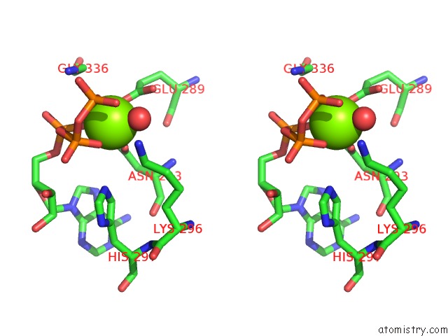
Stereo pair view
Mono view
Stereo pair view
A full contact list of Magnesium with other atoms in the Mg binding
site number 1 of Crystal Structure of the Atp-Binding Domain of Desk in Complex with Atp within 5.0Å range:
|
Reference:
F.Trajtenberg,
M.Grana,
N.Ruetalo,
H.Botti,
A.Buschiazzo.
Structural and Enzymatic Insights Into the Atp Binding and Autophosphorylation Mechanism of A Sensor Histidine Kinase J.Biol.Chem. V. 285 24892 2010.
ISSN: ISSN 0021-9258
PubMed: 20507988
DOI: 10.1074/JBC.M110.147843
Page generated: Wed Aug 14 13:04:11 2024
ISSN: ISSN 0021-9258
PubMed: 20507988
DOI: 10.1074/JBC.M110.147843
Last articles
Zn in 9JYWZn in 9IR4
Zn in 9IR3
Zn in 9GMX
Zn in 9GMW
Zn in 9JEJ
Zn in 9ERF
Zn in 9ERE
Zn in 9EGV
Zn in 9EGW