Magnesium »
PDB 3efq-3eql »
3eps »
Magnesium in PDB 3eps: The Crystal Structure of Isocitrate Dehydrogenase Kinase/Phosphatase From E. Coli
Enzymatic activity of The Crystal Structure of Isocitrate Dehydrogenase Kinase/Phosphatase From E. Coli
All present enzymatic activity of The Crystal Structure of Isocitrate Dehydrogenase Kinase/Phosphatase From E. Coli:
2.7.11.5;
2.7.11.5;
Protein crystallography data
The structure of The Crystal Structure of Isocitrate Dehydrogenase Kinase/Phosphatase From E. Coli, PDB code: 3eps
was solved by
J.Zheng,
Z.Jia,
Montreal-Kingston Bacterial Structural Genomicsinitiative (Bsgi),
with X-Ray Crystallography technique. A brief refinement statistics is given in the table below:
| Resolution Low / High (Å) | 30.00 / 2.80 |
| Space group | P 41 21 2 |
| Cell size a, b, c (Å), α, β, γ (°) | 124.595, 124.595, 267.635, 90.00, 90.00, 90.00 |
| R / Rfree (%) | 23.3 / 26.8 |
Magnesium Binding Sites:
The binding sites of Magnesium atom in the The Crystal Structure of Isocitrate Dehydrogenase Kinase/Phosphatase From E. Coli
(pdb code 3eps). This binding sites where shown within
5.0 Angstroms radius around Magnesium atom.
In total 2 binding sites of Magnesium where determined in the The Crystal Structure of Isocitrate Dehydrogenase Kinase/Phosphatase From E. Coli, PDB code: 3eps:
Jump to Magnesium binding site number: 1; 2;
In total 2 binding sites of Magnesium where determined in the The Crystal Structure of Isocitrate Dehydrogenase Kinase/Phosphatase From E. Coli, PDB code: 3eps:
Jump to Magnesium binding site number: 1; 2;
Magnesium binding site 1 out of 2 in 3eps
Go back to
Magnesium binding site 1 out
of 2 in the The Crystal Structure of Isocitrate Dehydrogenase Kinase/Phosphatase From E. Coli
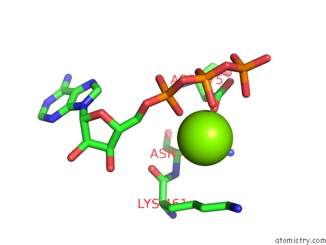
Mono view
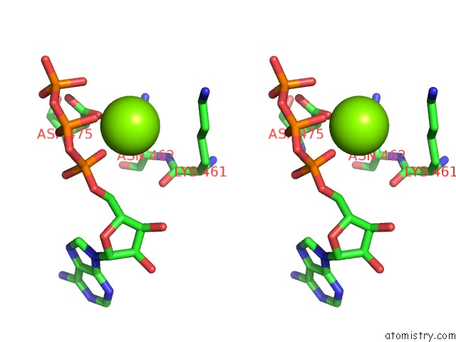
Stereo pair view
Mono view
Stereo pair view
A full contact list of Magnesium with other atoms in the Mg binding
site number 1 of The Crystal Structure of Isocitrate Dehydrogenase Kinase/Phosphatase From E. Coli within 5.0Å range:
|
Magnesium binding site 2 out of 2 in 3eps
Go back to
Magnesium binding site 2 out
of 2 in the The Crystal Structure of Isocitrate Dehydrogenase Kinase/Phosphatase From E. Coli
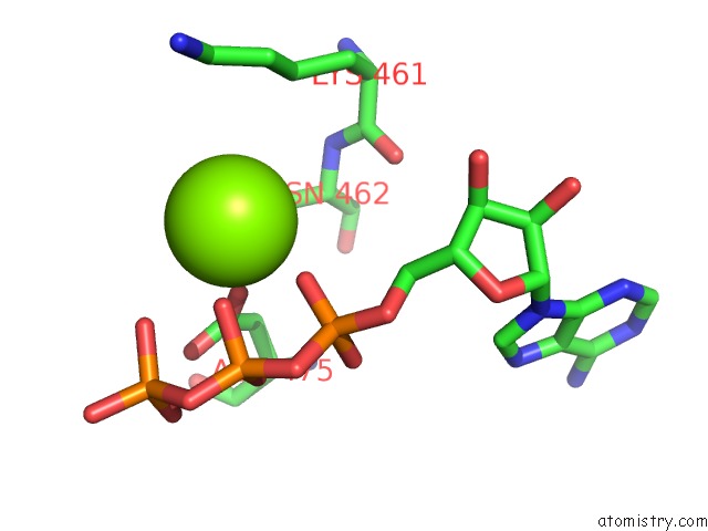
Mono view
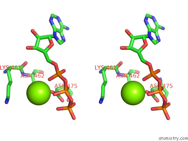
Stereo pair view
Mono view
Stereo pair view
A full contact list of Magnesium with other atoms in the Mg binding
site number 2 of The Crystal Structure of Isocitrate Dehydrogenase Kinase/Phosphatase From E. Coli within 5.0Å range:
|
Reference:
J.Zheng,
Z.Jia.
Structure of the Bifunctional Isocitrate Dehydrogenase Kinase/Phosphatase. Nature V. 465 961 2010.
ISSN: ISSN 0028-0836
PubMed: 20505668
DOI: 10.1038/NATURE09088
Page generated: Wed Aug 14 13:14:49 2024
ISSN: ISSN 0028-0836
PubMed: 20505668
DOI: 10.1038/NATURE09088
Last articles
Zn in 9JYWZn in 9IR4
Zn in 9IR3
Zn in 9GMX
Zn in 9GMW
Zn in 9JEJ
Zn in 9ERF
Zn in 9ERE
Zn in 9EGV
Zn in 9EGW