Magnesium »
PDB 3es7-3f2q »
3es8 »
Magnesium in PDB 3es8: Crystal Structure of Divergent Enolase From Oceanobacillus Iheyensis Complexed with Mg and L-Malate.
Protein crystallography data
The structure of Crystal Structure of Divergent Enolase From Oceanobacillus Iheyensis Complexed with Mg and L-Malate., PDB code: 3es8
was solved by
A.A.Fedorov,
E.V.Fedorov,
J.M.Sauder,
S.K.Burley,
J.A.Gerlt,
S.C.Almo,
Newyork Sgx Research Center For Structural Genomics (Nysgxrc),
with X-Ray Crystallography technique. A brief refinement statistics is given in the table below:
| Resolution Low / High (Å) | 24.94 / 2.20 |
| Space group | P 1 |
| Cell size a, b, c (Å), α, β, γ (°) | 105.584, 105.695, 105.718, 109.31, 109.47, 109.66 |
| R / Rfree (%) | 22.4 / 24.1 |
Magnesium Binding Sites:
The binding sites of Magnesium atom in the Crystal Structure of Divergent Enolase From Oceanobacillus Iheyensis Complexed with Mg and L-Malate.
(pdb code 3es8). This binding sites where shown within
5.0 Angstroms radius around Magnesium atom.
In total 8 binding sites of Magnesium where determined in the Crystal Structure of Divergent Enolase From Oceanobacillus Iheyensis Complexed with Mg and L-Malate., PDB code: 3es8:
Jump to Magnesium binding site number: 1; 2; 3; 4; 5; 6; 7; 8;
In total 8 binding sites of Magnesium where determined in the Crystal Structure of Divergent Enolase From Oceanobacillus Iheyensis Complexed with Mg and L-Malate., PDB code: 3es8:
Jump to Magnesium binding site number: 1; 2; 3; 4; 5; 6; 7; 8;
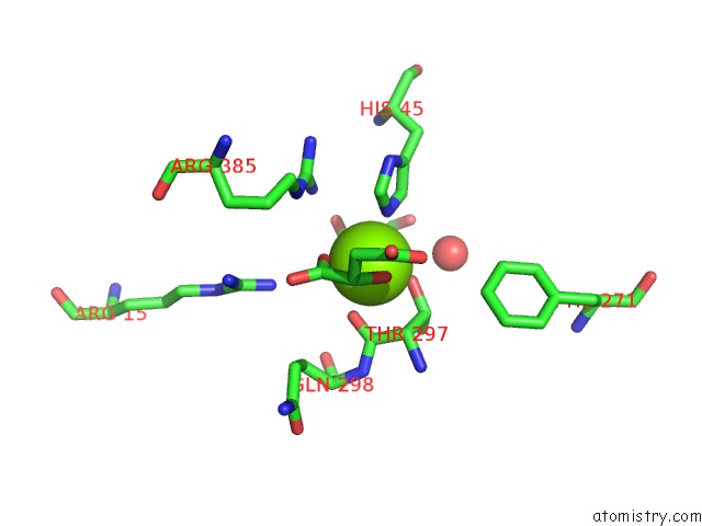
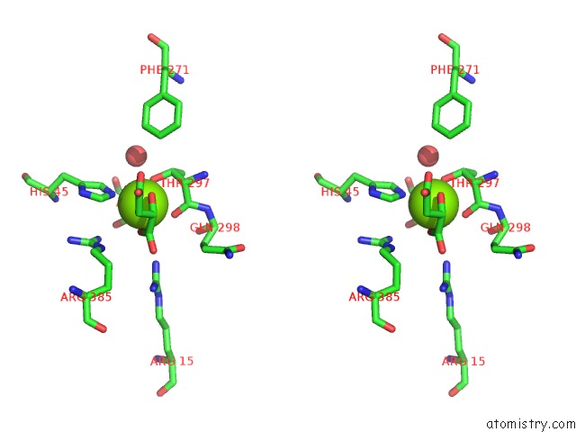
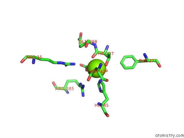
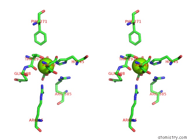
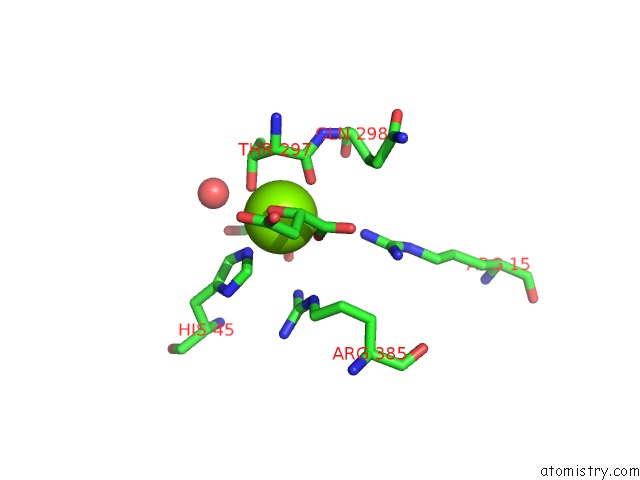
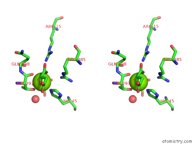
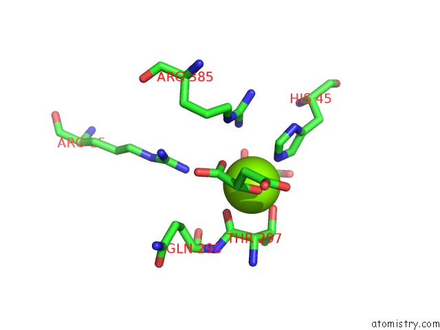
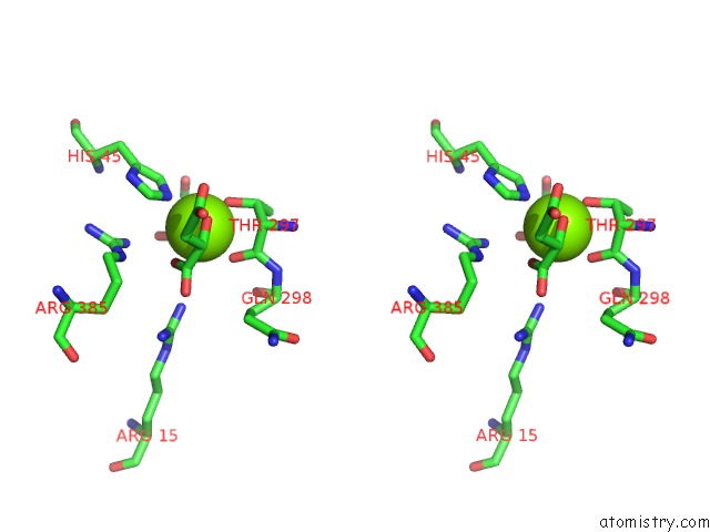
Magnesium binding site 1 out of 8 in 3es8
Go back to
Magnesium binding site 1 out
of 8 in the Crystal Structure of Divergent Enolase From Oceanobacillus Iheyensis Complexed with Mg and L-Malate.
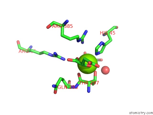
Mono view
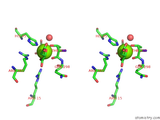
Stereo pair view
Mono view
Stereo pair view
A full contact list of Magnesium with other atoms in the Mg binding
site number 1 of Crystal Structure of Divergent Enolase From Oceanobacillus Iheyensis Complexed with Mg and L-Malate. within 5.0Å range:
|
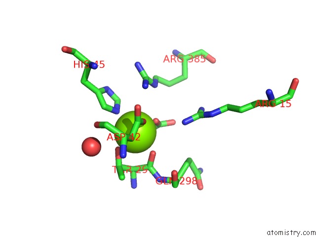
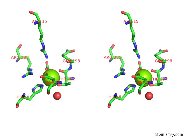
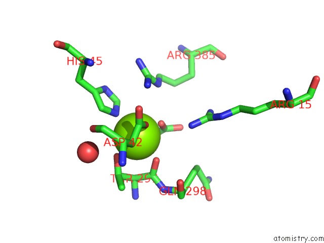
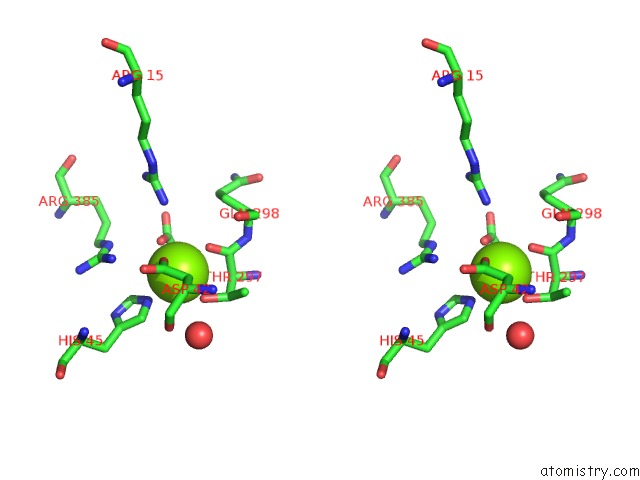
Magnesium binding site 2 out of 8 in 3es8
Go back to
Magnesium binding site 2 out
of 8 in the Crystal Structure of Divergent Enolase From Oceanobacillus Iheyensis Complexed with Mg and L-Malate.
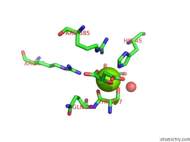
Mono view
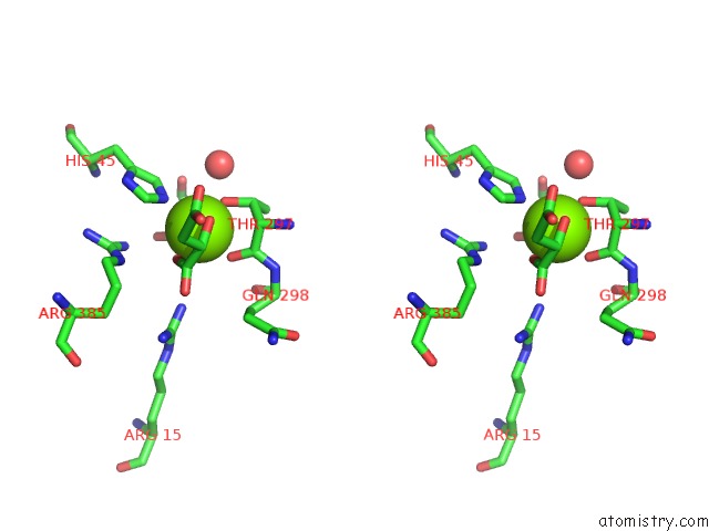
Stereo pair view
Mono view
Stereo pair view
A full contact list of Magnesium with other atoms in the Mg binding
site number 2 of Crystal Structure of Divergent Enolase From Oceanobacillus Iheyensis Complexed with Mg and L-Malate. within 5.0Å range:
|
Magnesium binding site 3 out of 8 in 3es8
Go back to
Magnesium binding site 3 out
of 8 in the Crystal Structure of Divergent Enolase From Oceanobacillus Iheyensis Complexed with Mg and L-Malate.
Mono view
Stereo pair view
Mono view
Stereo pair view
A full contact list of Magnesium with other atoms in the Mg binding
site number 3 of Crystal Structure of Divergent Enolase From Oceanobacillus Iheyensis Complexed with Mg and L-Malate. within 5.0Å range:
|
Magnesium binding site 4 out of 8 in 3es8
Go back to
Magnesium binding site 4 out
of 8 in the Crystal Structure of Divergent Enolase From Oceanobacillus Iheyensis Complexed with Mg and L-Malate.
Mono view
Stereo pair view
Mono view
Stereo pair view
A full contact list of Magnesium with other atoms in the Mg binding
site number 4 of Crystal Structure of Divergent Enolase From Oceanobacillus Iheyensis Complexed with Mg and L-Malate. within 5.0Å range:
|
Magnesium binding site 5 out of 8 in 3es8
Go back to
Magnesium binding site 5 out
of 8 in the Crystal Structure of Divergent Enolase From Oceanobacillus Iheyensis Complexed with Mg and L-Malate.
Mono view
Stereo pair view
Mono view
Stereo pair view
A full contact list of Magnesium with other atoms in the Mg binding
site number 5 of Crystal Structure of Divergent Enolase From Oceanobacillus Iheyensis Complexed with Mg and L-Malate. within 5.0Å range:
|
Magnesium binding site 6 out of 8 in 3es8
Go back to
Magnesium binding site 6 out
of 8 in the Crystal Structure of Divergent Enolase From Oceanobacillus Iheyensis Complexed with Mg and L-Malate.
Mono view
Stereo pair view
Mono view
Stereo pair view
A full contact list of Magnesium with other atoms in the Mg binding
site number 6 of Crystal Structure of Divergent Enolase From Oceanobacillus Iheyensis Complexed with Mg and L-Malate. within 5.0Å range:
|
Magnesium binding site 7 out of 8 in 3es8
Go back to
Magnesium binding site 7 out
of 8 in the Crystal Structure of Divergent Enolase From Oceanobacillus Iheyensis Complexed with Mg and L-Malate.
Mono view
Stereo pair view
Mono view
Stereo pair view
A full contact list of Magnesium with other atoms in the Mg binding
site number 7 of Crystal Structure of Divergent Enolase From Oceanobacillus Iheyensis Complexed with Mg and L-Malate. within 5.0Å range:
|
Magnesium binding site 8 out of 8 in 3es8
Go back to
Magnesium binding site 8 out
of 8 in the Crystal Structure of Divergent Enolase From Oceanobacillus Iheyensis Complexed with Mg and L-Malate.
Mono view
Stereo pair view
Mono view
Stereo pair view
A full contact list of Magnesium with other atoms in the Mg binding
site number 8 of Crystal Structure of Divergent Enolase From Oceanobacillus Iheyensis Complexed with Mg and L-Malate. within 5.0Å range:
|
Reference:
J.F.Rakus,
C.Kalyanaraman,
A.A.Fedorov,
E.V.Fedorov,
F.P.Mills-Groninger,
R.Toro,
J.Bonanno,
K.Bain,
J.M.Sauder,
S.K.Burley,
S.C.Almo,
M.P.Jacobson,
J.A.Gerlt.
Computation-Facilitated Assignment of the Function in the Enolase Superfamily: A Regiochemically Distinct Galactarate Dehydratase From Oceanobacillus Iheyensis . Biochemistry V. 48 11546 2009.
ISSN: ISSN 0006-2960
PubMed: 19883118
DOI: 10.1021/BI901731C
Page generated: Wed Aug 14 13:19:50 2024
ISSN: ISSN 0006-2960
PubMed: 19883118
DOI: 10.1021/BI901731C
Last articles
Zn in 9JYWZn in 9IR4
Zn in 9IR3
Zn in 9GMX
Zn in 9GMW
Zn in 9JEJ
Zn in 9ERF
Zn in 9ERE
Zn in 9EGV
Zn in 9EGW