Magnesium »
PDB 3es7-3f2q »
3ey9 »
Magnesium in PDB 3ey9: Structural Basis For Membrane Binding and Catalytic Activation of the Peripheral Membrane Enzyme Pyruvate Oxidase From Escherichia Coli
Enzymatic activity of Structural Basis For Membrane Binding and Catalytic Activation of the Peripheral Membrane Enzyme Pyruvate Oxidase From Escherichia Coli
All present enzymatic activity of Structural Basis For Membrane Binding and Catalytic Activation of the Peripheral Membrane Enzyme Pyruvate Oxidase From Escherichia Coli:
1.2.2.2;
1.2.2.2;
Protein crystallography data
The structure of Structural Basis For Membrane Binding and Catalytic Activation of the Peripheral Membrane Enzyme Pyruvate Oxidase From Escherichia Coli, PDB code: 3ey9
was solved by
P.Neumann,
A.Weidner,
A.Pech,
M.T.Stubbs,
K.Tittmann,
with X-Ray Crystallography technique. A brief refinement statistics is given in the table below:
| Resolution Low / High (Å) | 29.97 / 2.90 |
| Space group | P 43 21 2 |
| Cell size a, b, c (Å), α, β, γ (°) | 151.370, 151.370, 153.740, 90.00, 90.00, 90.00 |
| R / Rfree (%) | 18.2 / 21.6 |
Magnesium Binding Sites:
The binding sites of Magnesium atom in the Structural Basis For Membrane Binding and Catalytic Activation of the Peripheral Membrane Enzyme Pyruvate Oxidase From Escherichia Coli
(pdb code 3ey9). This binding sites where shown within
5.0 Angstroms radius around Magnesium atom.
In total 2 binding sites of Magnesium where determined in the Structural Basis For Membrane Binding and Catalytic Activation of the Peripheral Membrane Enzyme Pyruvate Oxidase From Escherichia Coli, PDB code: 3ey9:
Jump to Magnesium binding site number: 1; 2;
In total 2 binding sites of Magnesium where determined in the Structural Basis For Membrane Binding and Catalytic Activation of the Peripheral Membrane Enzyme Pyruvate Oxidase From Escherichia Coli, PDB code: 3ey9:
Jump to Magnesium binding site number: 1; 2;
Magnesium binding site 1 out of 2 in 3ey9
Go back to
Magnesium binding site 1 out
of 2 in the Structural Basis For Membrane Binding and Catalytic Activation of the Peripheral Membrane Enzyme Pyruvate Oxidase From Escherichia Coli
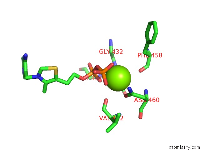
Mono view
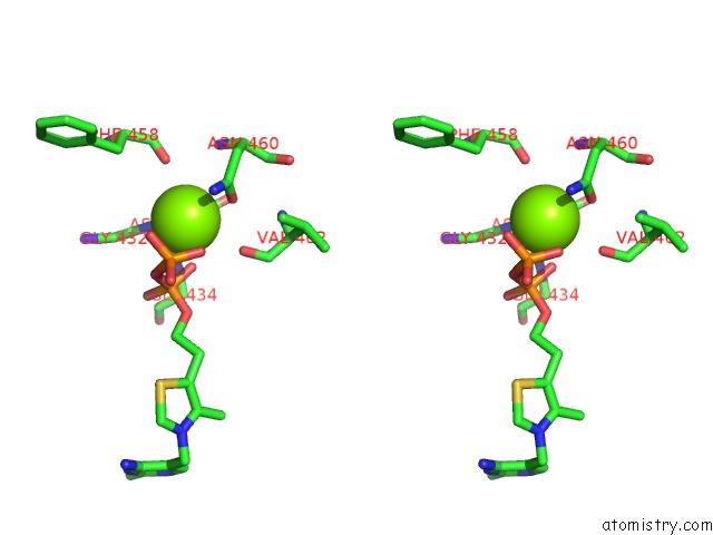
Stereo pair view
Mono view
Stereo pair view
A full contact list of Magnesium with other atoms in the Mg binding
site number 1 of Structural Basis For Membrane Binding and Catalytic Activation of the Peripheral Membrane Enzyme Pyruvate Oxidase From Escherichia Coli within 5.0Å range:
|
Magnesium binding site 2 out of 2 in 3ey9
Go back to
Magnesium binding site 2 out
of 2 in the Structural Basis For Membrane Binding and Catalytic Activation of the Peripheral Membrane Enzyme Pyruvate Oxidase From Escherichia Coli
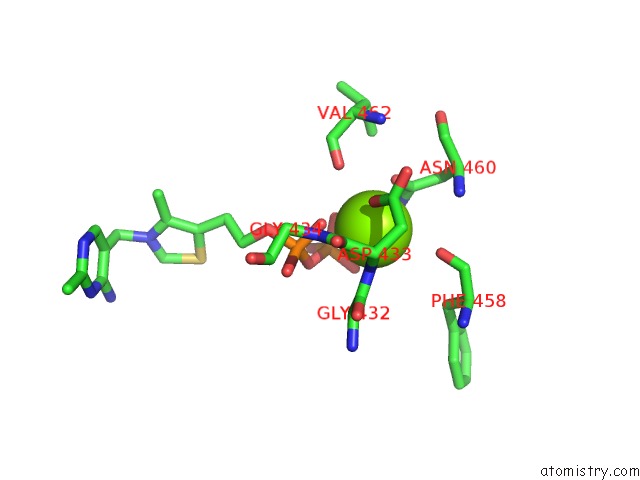
Mono view
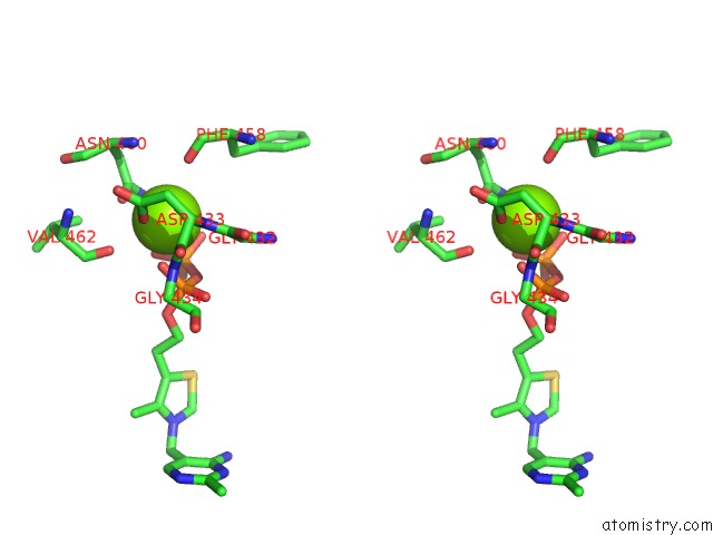
Stereo pair view
Mono view
Stereo pair view
A full contact list of Magnesium with other atoms in the Mg binding
site number 2 of Structural Basis For Membrane Binding and Catalytic Activation of the Peripheral Membrane Enzyme Pyruvate Oxidase From Escherichia Coli within 5.0Å range:
|
Reference:
P.Neumann,
A.Weidner,
A.Pech,
M.T.Stubbs,
K.Tittmann.
Structural Basis For Membrane Binding and Catalytic Activation of the Peripheral Membrane Enzyme Pyruvate Oxidase From Escherichia Coli. Proc.Natl.Acad.Sci.Usa V. 105 17390 2008.
ISSN: ISSN 0027-8424
PubMed: 18988747
DOI: 10.1073/PNAS.0805027105
Page generated: Wed Aug 14 13:23:17 2024
ISSN: ISSN 0027-8424
PubMed: 18988747
DOI: 10.1073/PNAS.0805027105
Last articles
Zn in 9MJ5Zn in 9HNW
Zn in 9G0L
Zn in 9FNE
Zn in 9DZN
Zn in 9E0I
Zn in 9D32
Zn in 9DAK
Zn in 8ZXC
Zn in 8ZUF