Magnesium »
PDB 3f2t-3fcv »
3f4e »
Magnesium in PDB 3f4e: Crystal Structure of the Fmn Riboswitch Bound to Fmn, Split Rna.
Protein crystallography data
The structure of Crystal Structure of the Fmn Riboswitch Bound to Fmn, Split Rna., PDB code: 3f4e
was solved by
A.A.Serganov,
L.Huang,
with X-Ray Crystallography technique. A brief refinement statistics is given in the table below:
| Resolution Low / High (Å) | 20.00 / 3.05 |
| Space group | P 31 2 1 |
| Cell size a, b, c (Å), α, β, γ (°) | 71.471, 71.471, 140.554, 90.00, 90.00, 120.00 |
| R / Rfree (%) | 20 / 23.2 |
Other elements in 3f4e:
The structure of Crystal Structure of the Fmn Riboswitch Bound to Fmn, Split Rna. also contains other interesting chemical elements:
| Potassium | (K) | 1 atom |
Magnesium Binding Sites:
Pages:
>>> Page 1 <<< Page 2, Binding sites: 11 - 13;Binding sites:
The binding sites of Magnesium atom in the Crystal Structure of the Fmn Riboswitch Bound to Fmn, Split Rna. (pdb code 3f4e). This binding sites where shown within 5.0 Angstroms radius around Magnesium atom.In total 13 binding sites of Magnesium where determined in the Crystal Structure of the Fmn Riboswitch Bound to Fmn, Split Rna., PDB code: 3f4e:
Jump to Magnesium binding site number: 1; 2; 3; 4; 5; 6; 7; 8; 9; 10;
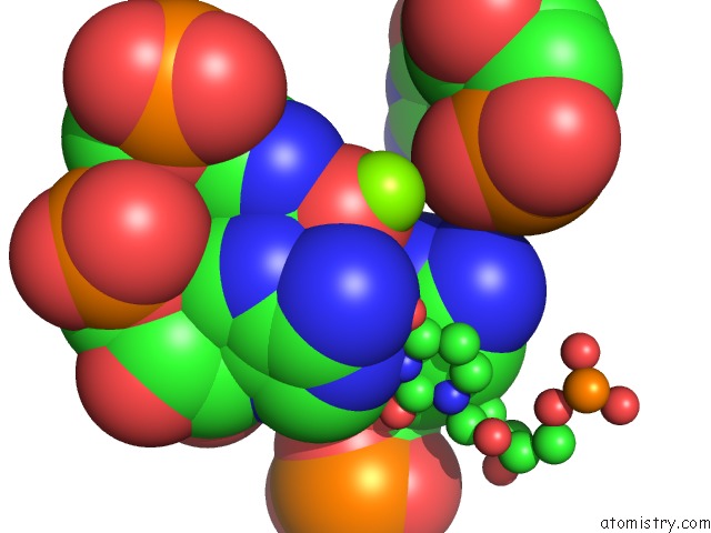
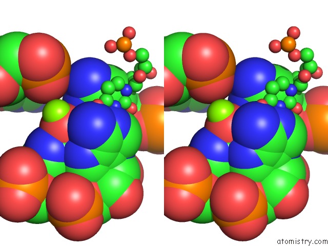
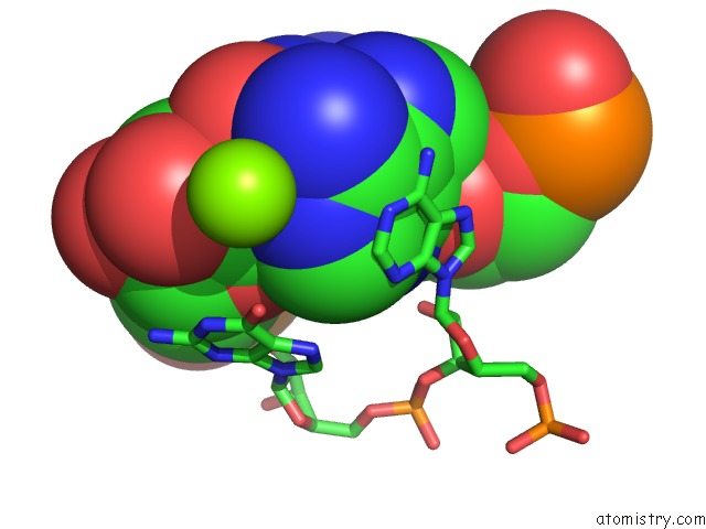
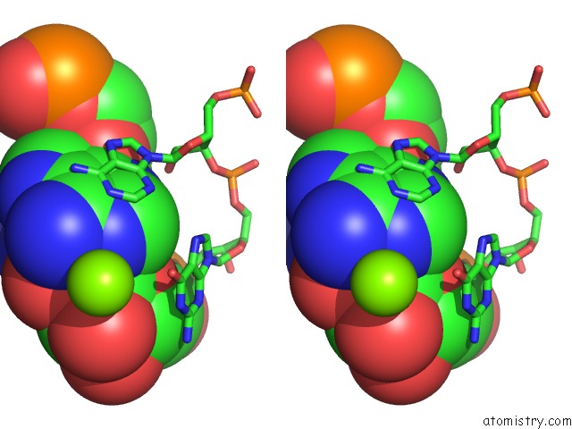
Magnesium binding site 1 out of 13 in 3f4e
Go back to
Magnesium binding site 1 out
of 13 in the Crystal Structure of the Fmn Riboswitch Bound to Fmn, Split Rna.
Mono view
Stereo pair view
Mono view
Stereo pair view
A full contact list of Magnesium with other atoms in the Mg binding
site number 1 of Crystal Structure of the Fmn Riboswitch Bound to Fmn, Split Rna. within 5.0Å range:
|
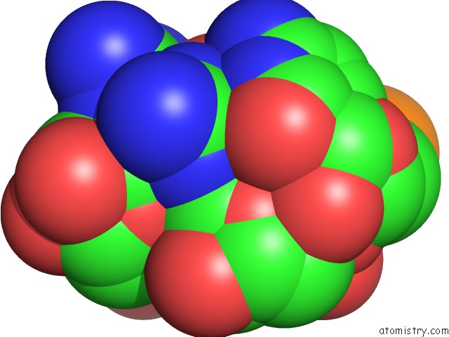
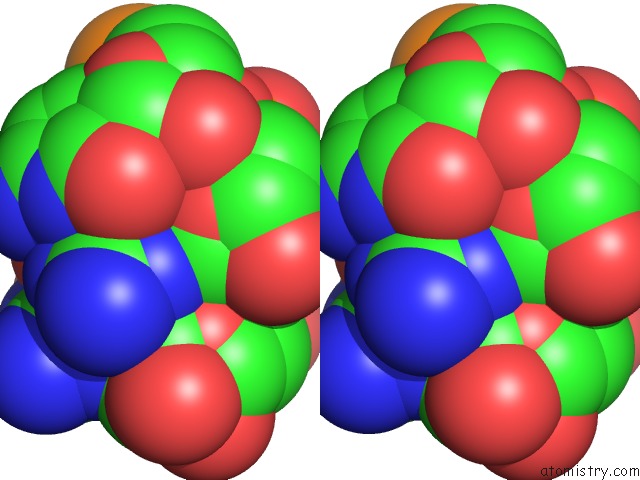
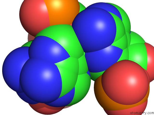
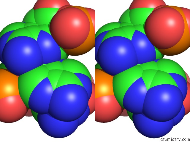
Magnesium binding site 2 out of 13 in 3f4e
Go back to
Magnesium binding site 2 out
of 13 in the Crystal Structure of the Fmn Riboswitch Bound to Fmn, Split Rna.
Mono view
Stereo pair view
Mono view
Stereo pair view
A full contact list of Magnesium with other atoms in the Mg binding
site number 2 of Crystal Structure of the Fmn Riboswitch Bound to Fmn, Split Rna. within 5.0Å range:
|
Magnesium binding site 3 out of 13 in 3f4e
Go back to
Magnesium binding site 3 out
of 13 in the Crystal Structure of the Fmn Riboswitch Bound to Fmn, Split Rna.
Mono view
Stereo pair view
Mono view
Stereo pair view
A full contact list of Magnesium with other atoms in the Mg binding
site number 3 of Crystal Structure of the Fmn Riboswitch Bound to Fmn, Split Rna. within 5.0Å range:
|
Magnesium binding site 4 out of 13 in 3f4e
Go back to
Magnesium binding site 4 out
of 13 in the Crystal Structure of the Fmn Riboswitch Bound to Fmn, Split Rna.
Mono view
Stereo pair view
Mono view
Stereo pair view
A full contact list of Magnesium with other atoms in the Mg binding
site number 4 of Crystal Structure of the Fmn Riboswitch Bound to Fmn, Split Rna. within 5.0Å range:
|
Magnesium binding site 5 out of 13 in 3f4e
Go back to
Magnesium binding site 5 out
of 13 in the Crystal Structure of the Fmn Riboswitch Bound to Fmn, Split Rna.
Mono view
Stereo pair view
Mono view
Stereo pair view
A full contact list of Magnesium with other atoms in the Mg binding
site number 5 of Crystal Structure of the Fmn Riboswitch Bound to Fmn, Split Rna. within 5.0Å range:
|
Magnesium binding site 6 out of 13 in 3f4e
Go back to
Magnesium binding site 6 out
of 13 in the Crystal Structure of the Fmn Riboswitch Bound to Fmn, Split Rna.
Mono view
Stereo pair view
Mono view
Stereo pair view
A full contact list of Magnesium with other atoms in the Mg binding
site number 6 of Crystal Structure of the Fmn Riboswitch Bound to Fmn, Split Rna. within 5.0Å range:
|
Magnesium binding site 7 out of 13 in 3f4e
Go back to
Magnesium binding site 7 out
of 13 in the Crystal Structure of the Fmn Riboswitch Bound to Fmn, Split Rna.
Mono view
Stereo pair view
Mono view
Stereo pair view
A full contact list of Magnesium with other atoms in the Mg binding
site number 7 of Crystal Structure of the Fmn Riboswitch Bound to Fmn, Split Rna. within 5.0Å range:
|
Magnesium binding site 8 out of 13 in 3f4e
Go back to
Magnesium binding site 8 out
of 13 in the Crystal Structure of the Fmn Riboswitch Bound to Fmn, Split Rna.
Mono view
Stereo pair view
Mono view
Stereo pair view
A full contact list of Magnesium with other atoms in the Mg binding
site number 8 of Crystal Structure of the Fmn Riboswitch Bound to Fmn, Split Rna. within 5.0Å range:
|
Magnesium binding site 9 out of 13 in 3f4e
Go back to
Magnesium binding site 9 out
of 13 in the Crystal Structure of the Fmn Riboswitch Bound to Fmn, Split Rna.
Mono view
Stereo pair view
Mono view
Stereo pair view
A full contact list of Magnesium with other atoms in the Mg binding
site number 9 of Crystal Structure of the Fmn Riboswitch Bound to Fmn, Split Rna. within 5.0Å range:
|
Magnesium binding site 10 out of 13 in 3f4e
Go back to
Magnesium binding site 10 out
of 13 in the Crystal Structure of the Fmn Riboswitch Bound to Fmn, Split Rna.
Mono view
Stereo pair view
Mono view
Stereo pair view
A full contact list of Magnesium with other atoms in the Mg binding
site number 10 of Crystal Structure of the Fmn Riboswitch Bound to Fmn, Split Rna. within 5.0Å range:
|
Reference:
A.Serganov,
L.Huang,
D.J.Patel.
Coenzyme Recognition and Gene Regulation By A Flavin Mononucleotide Riboswitch. Nature V. 458 233 2009.
ISSN: ISSN 0028-0836
PubMed: 19169240
DOI: 10.1038/NATURE07642
Page generated: Wed Aug 14 13:28:49 2024
ISSN: ISSN 0028-0836
PubMed: 19169240
DOI: 10.1038/NATURE07642
Last articles
Zn in 9MJ5Zn in 9HNW
Zn in 9G0L
Zn in 9FNE
Zn in 9DZN
Zn in 9E0I
Zn in 9D32
Zn in 9DAK
Zn in 8ZXC
Zn in 8ZUF