Magnesium »
PDB 3f2t-3fcv »
3f61 »
Magnesium in PDB 3f61: Crystal Structure of M. Tuberculosis Pknb LEU33ASP/VAL222ASP Double Mutant in Complex with Adp
Enzymatic activity of Crystal Structure of M. Tuberculosis Pknb LEU33ASP/VAL222ASP Double Mutant in Complex with Adp
All present enzymatic activity of Crystal Structure of M. Tuberculosis Pknb LEU33ASP/VAL222ASP Double Mutant in Complex with Adp:
2.7.11.1;
2.7.11.1;
Protein crystallography data
The structure of Crystal Structure of M. Tuberculosis Pknb LEU33ASP/VAL222ASP Double Mutant in Complex with Adp, PDB code: 3f61
was solved by
C.A.Mieczkowski,
T.Alber,
Tb Structural Genomics Consortium (Tbsgc),
with X-Ray Crystallography technique. A brief refinement statistics is given in the table below:
| Resolution Low / High (Å) | 37.77 / 1.80 |
| Space group | P 21 21 21 |
| Cell size a, b, c (Å), α, β, γ (°) | 38.981, 51.542, 152.434, 90.00, 90.00, 90.00 |
| R / Rfree (%) | 20.3 / 22.8 |
Magnesium Binding Sites:
The binding sites of Magnesium atom in the Crystal Structure of M. Tuberculosis Pknb LEU33ASP/VAL222ASP Double Mutant in Complex with Adp
(pdb code 3f61). This binding sites where shown within
5.0 Angstroms radius around Magnesium atom.
In total 4 binding sites of Magnesium where determined in the Crystal Structure of M. Tuberculosis Pknb LEU33ASP/VAL222ASP Double Mutant in Complex with Adp, PDB code: 3f61:
Jump to Magnesium binding site number: 1; 2; 3; 4;
In total 4 binding sites of Magnesium where determined in the Crystal Structure of M. Tuberculosis Pknb LEU33ASP/VAL222ASP Double Mutant in Complex with Adp, PDB code: 3f61:
Jump to Magnesium binding site number: 1; 2; 3; 4;
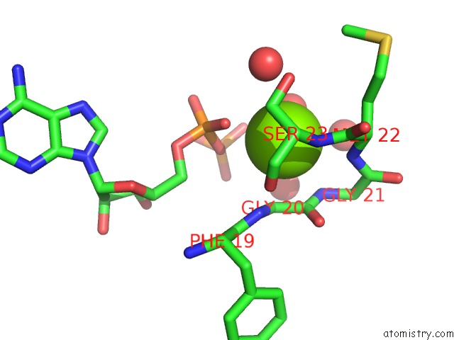
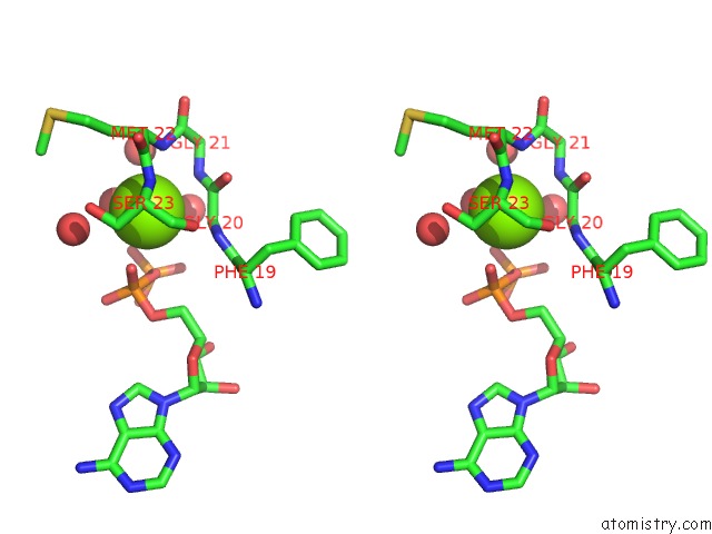
Magnesium binding site 1 out of 4 in 3f61
Go back to
Magnesium binding site 1 out
of 4 in the Crystal Structure of M. Tuberculosis Pknb LEU33ASP/VAL222ASP Double Mutant in Complex with Adp
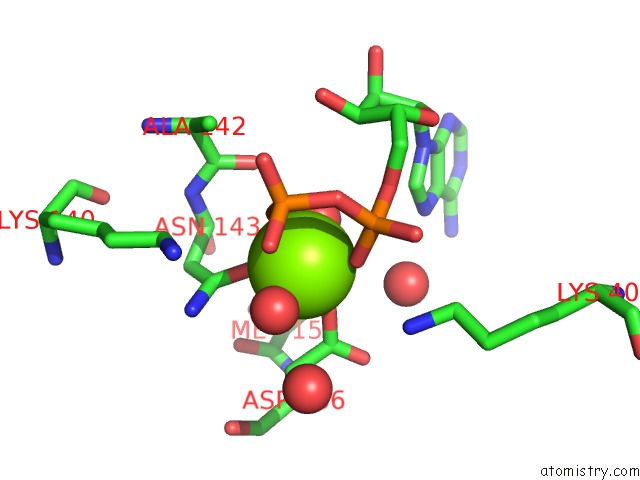
Mono view
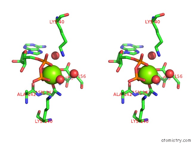
Stereo pair view
Mono view
Stereo pair view
A full contact list of Magnesium with other atoms in the Mg binding
site number 1 of Crystal Structure of M. Tuberculosis Pknb LEU33ASP/VAL222ASP Double Mutant in Complex with Adp within 5.0Å range:
|
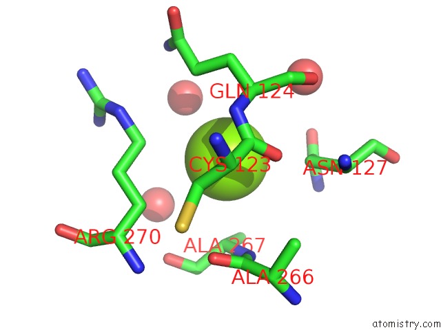
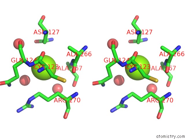
Magnesium binding site 2 out of 4 in 3f61
Go back to
Magnesium binding site 2 out
of 4 in the Crystal Structure of M. Tuberculosis Pknb LEU33ASP/VAL222ASP Double Mutant in Complex with Adp
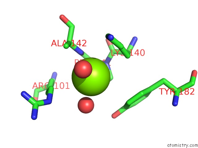
Mono view
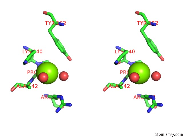
Stereo pair view
Mono view
Stereo pair view
A full contact list of Magnesium with other atoms in the Mg binding
site number 2 of Crystal Structure of M. Tuberculosis Pknb LEU33ASP/VAL222ASP Double Mutant in Complex with Adp within 5.0Å range:
|
Magnesium binding site 3 out of 4 in 3f61
Go back to
Magnesium binding site 3 out
of 4 in the Crystal Structure of M. Tuberculosis Pknb LEU33ASP/VAL222ASP Double Mutant in Complex with Adp
Mono view
Stereo pair view
Mono view
Stereo pair view
A full contact list of Magnesium with other atoms in the Mg binding
site number 3 of Crystal Structure of M. Tuberculosis Pknb LEU33ASP/VAL222ASP Double Mutant in Complex with Adp within 5.0Å range:
|
Magnesium binding site 4 out of 4 in 3f61
Go back to
Magnesium binding site 4 out
of 4 in the Crystal Structure of M. Tuberculosis Pknb LEU33ASP/VAL222ASP Double Mutant in Complex with Adp
Mono view
Stereo pair view
Mono view
Stereo pair view
A full contact list of Magnesium with other atoms in the Mg binding
site number 4 of Crystal Structure of M. Tuberculosis Pknb LEU33ASP/VAL222ASP Double Mutant in Complex with Adp within 5.0Å range:
|
Reference:
C.Mieczkowski,
A.T.Iavarone,
T.Alber.
Auto-Activation Mechanism of the Mycobacterium Tuberculosis Pknb Receptor Ser/Thr Kinase. Embo J. V. 27 3186 2008.
ISSN: ISSN 0261-4189
PubMed: 19008858
DOI: 10.1038/EMBOJ.2008.236
Page generated: Wed Aug 14 13:33:59 2024
ISSN: ISSN 0261-4189
PubMed: 19008858
DOI: 10.1038/EMBOJ.2008.236
Last articles
Cl in 5Z5CCl in 5Z6Q
Cl in 5Z5Y
Cl in 5Z5B
Cl in 5Z5O
Cl in 5Z42
Cl in 5Z30
Cl in 5YZ8
Cl in 5Z2B
Cl in 5Z1Q