Magnesium »
PDB 3f2t-3fcv »
3f6e »
Magnesium in PDB 3f6e: Crystal Structure of Benzoylformate Decarboxylase in Complex with the Pyridyl Inhibitor 3-Pkb
Enzymatic activity of Crystal Structure of Benzoylformate Decarboxylase in Complex with the Pyridyl Inhibitor 3-Pkb
All present enzymatic activity of Crystal Structure of Benzoylformate Decarboxylase in Complex with the Pyridyl Inhibitor 3-Pkb:
4.1.1.7;
4.1.1.7;
Protein crystallography data
The structure of Crystal Structure of Benzoylformate Decarboxylase in Complex with the Pyridyl Inhibitor 3-Pkb, PDB code: 3f6e
was solved by
G.S.Brandt,
M.J.Mcleish,
G.L.Kenyon,
G.A.Petsko,
D.Ringe,
F.Jordan,
with X-Ray Crystallography technique. A brief refinement statistics is given in the table below:
| Resolution Low / High (Å) | 41.31 / 1.34 |
| Space group | I 2 2 2 |
| Cell size a, b, c (Å), α, β, γ (°) | 81.090, 95.807, 137.324, 90.00, 90.00, 90.00 |
| R / Rfree (%) | 19 / 21.2 |
Magnesium Binding Sites:
The binding sites of Magnesium atom in the Crystal Structure of Benzoylformate Decarboxylase in Complex with the Pyridyl Inhibitor 3-Pkb
(pdb code 3f6e). This binding sites where shown within
5.0 Angstroms radius around Magnesium atom.
In total only one binding site of Magnesium was determined in the Crystal Structure of Benzoylformate Decarboxylase in Complex with the Pyridyl Inhibitor 3-Pkb, PDB code: 3f6e:
In total only one binding site of Magnesium was determined in the Crystal Structure of Benzoylformate Decarboxylase in Complex with the Pyridyl Inhibitor 3-Pkb, PDB code: 3f6e:
Magnesium binding site 1 out of 1 in 3f6e
Go back to
Magnesium binding site 1 out
of 1 in the Crystal Structure of Benzoylformate Decarboxylase in Complex with the Pyridyl Inhibitor 3-Pkb
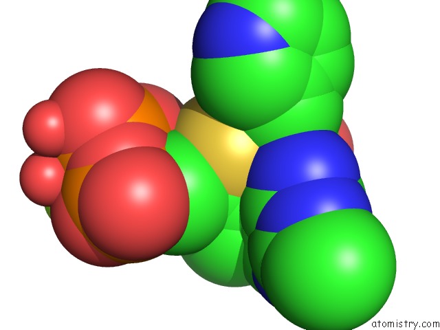
Mono view
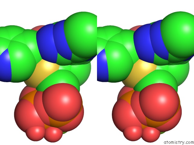
Stereo pair view
Mono view
Stereo pair view
A full contact list of Magnesium with other atoms in the Mg binding
site number 1 of Crystal Structure of Benzoylformate Decarboxylase in Complex with the Pyridyl Inhibitor 3-Pkb within 5.0Å range:
|
Reference:
S.Chakraborty,
N.S.Nemeria,
A.Balakrishnan,
G.S.Brandt,
M.M.Kneen,
A.Yep,
M.J.Mcleish,
G.L.Kenyon,
G.A.Petsko,
D.Ringe,
F.Jordan.
Detection and Time Course of Formation of Major Thiamin Diphosphate-Bound Covalent Intermediates Derived From A Chromophoric Substrate Analogue on Benzoylformate Decarboxylase. Biochemistry V. 48 981 2009.
ISSN: ISSN 0006-2960
PubMed: 19140682
DOI: 10.1021/BI801810H
Page generated: Wed Aug 14 13:34:22 2024
ISSN: ISSN 0006-2960
PubMed: 19140682
DOI: 10.1021/BI801810H
Last articles
Zn in 9J0NZn in 9J0O
Zn in 9J0P
Zn in 9FJX
Zn in 9EKB
Zn in 9C0F
Zn in 9CAH
Zn in 9CH0
Zn in 9CH3
Zn in 9CH1