Magnesium »
PDB 3f2t-3fcv »
3fbf »
Magnesium in PDB 3fbf: Crystal Structure of the Mimivirus Ndk N62L Mutant Complexed with Dtdp
Enzymatic activity of Crystal Structure of the Mimivirus Ndk N62L Mutant Complexed with Dtdp
All present enzymatic activity of Crystal Structure of the Mimivirus Ndk N62L Mutant Complexed with Dtdp:
2.7.4.6;
2.7.4.6;
Protein crystallography data
The structure of Crystal Structure of the Mimivirus Ndk N62L Mutant Complexed with Dtdp, PDB code: 3fbf
was solved by
S.Jeudy,
A.Lartigue,
J.M.Claverie,
C.Abergel,
with X-Ray Crystallography technique. A brief refinement statistics is given in the table below:
| Resolution Low / High (Å) | 28.62 / 2.60 |
| Space group | C 2 2 21 |
| Cell size a, b, c (Å), α, β, γ (°) | 80.241, 152.680, 185.248, 90.00, 90.00, 90.00 |
| R / Rfree (%) | 17.8 / 22.4 |
Magnesium Binding Sites:
The binding sites of Magnesium atom in the Crystal Structure of the Mimivirus Ndk N62L Mutant Complexed with Dtdp
(pdb code 3fbf). This binding sites where shown within
5.0 Angstroms radius around Magnesium atom.
In total 6 binding sites of Magnesium where determined in the Crystal Structure of the Mimivirus Ndk N62L Mutant Complexed with Dtdp, PDB code: 3fbf:
Jump to Magnesium binding site number: 1; 2; 3; 4; 5; 6;
In total 6 binding sites of Magnesium where determined in the Crystal Structure of the Mimivirus Ndk N62L Mutant Complexed with Dtdp, PDB code: 3fbf:
Jump to Magnesium binding site number: 1; 2; 3; 4; 5; 6;
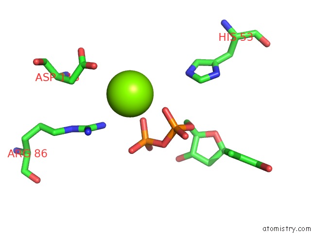
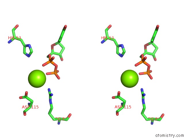
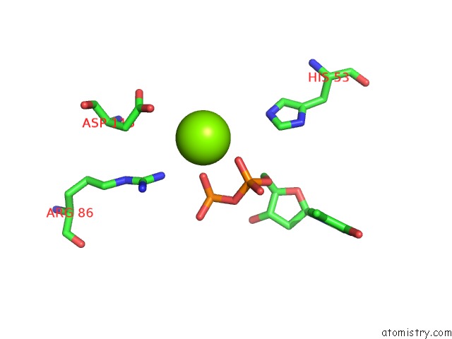
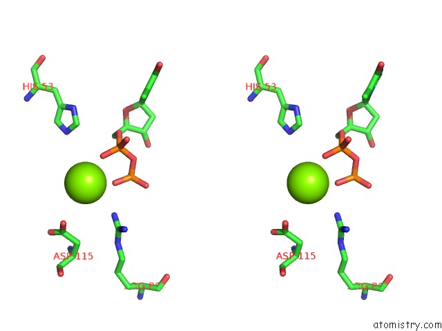
Magnesium binding site 1 out of 6 in 3fbf
Go back to
Magnesium binding site 1 out
of 6 in the Crystal Structure of the Mimivirus Ndk N62L Mutant Complexed with Dtdp
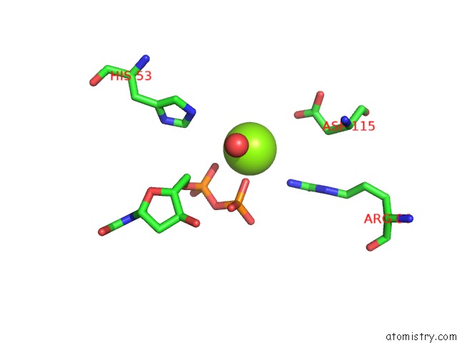
Mono view
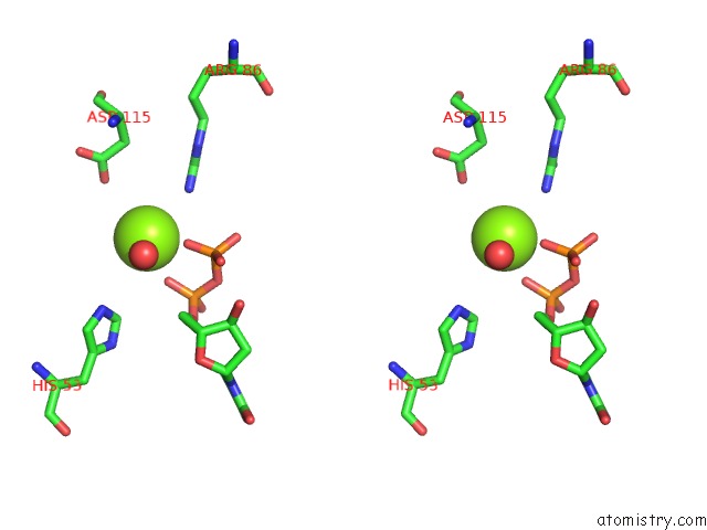
Stereo pair view
Mono view
Stereo pair view
A full contact list of Magnesium with other atoms in the Mg binding
site number 1 of Crystal Structure of the Mimivirus Ndk N62L Mutant Complexed with Dtdp within 5.0Å range:
|
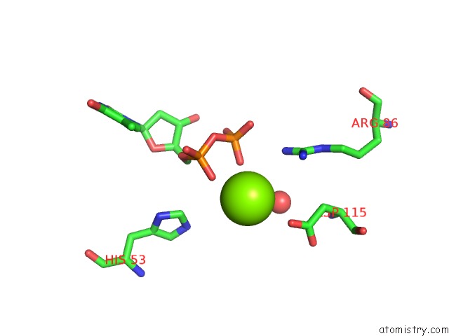
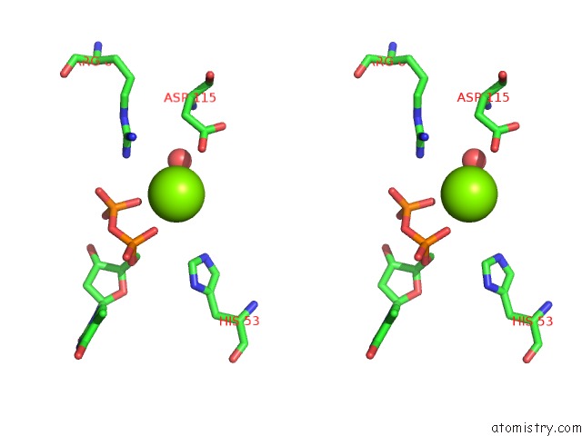
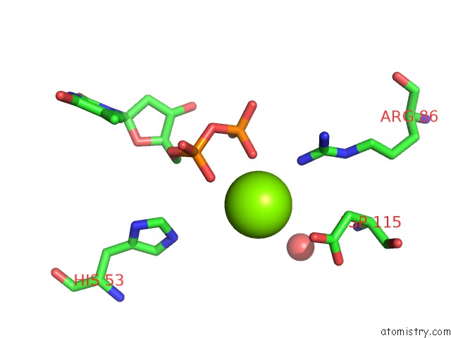
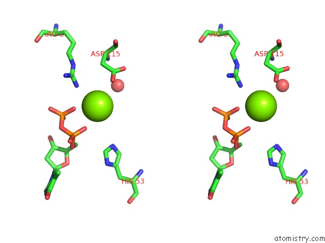
Magnesium binding site 2 out of 6 in 3fbf
Go back to
Magnesium binding site 2 out
of 6 in the Crystal Structure of the Mimivirus Ndk N62L Mutant Complexed with Dtdp
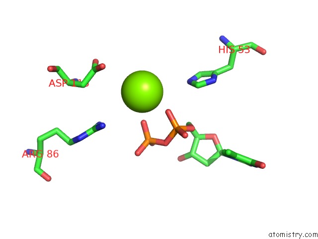
Mono view
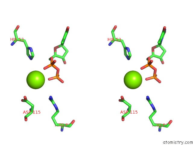
Stereo pair view
Mono view
Stereo pair view
A full contact list of Magnesium with other atoms in the Mg binding
site number 2 of Crystal Structure of the Mimivirus Ndk N62L Mutant Complexed with Dtdp within 5.0Å range:
|
Magnesium binding site 3 out of 6 in 3fbf
Go back to
Magnesium binding site 3 out
of 6 in the Crystal Structure of the Mimivirus Ndk N62L Mutant Complexed with Dtdp
Mono view
Stereo pair view
Mono view
Stereo pair view
A full contact list of Magnesium with other atoms in the Mg binding
site number 3 of Crystal Structure of the Mimivirus Ndk N62L Mutant Complexed with Dtdp within 5.0Å range:
|
Magnesium binding site 4 out of 6 in 3fbf
Go back to
Magnesium binding site 4 out
of 6 in the Crystal Structure of the Mimivirus Ndk N62L Mutant Complexed with Dtdp
Mono view
Stereo pair view
Mono view
Stereo pair view
A full contact list of Magnesium with other atoms in the Mg binding
site number 4 of Crystal Structure of the Mimivirus Ndk N62L Mutant Complexed with Dtdp within 5.0Å range:
|
Magnesium binding site 5 out of 6 in 3fbf
Go back to
Magnesium binding site 5 out
of 6 in the Crystal Structure of the Mimivirus Ndk N62L Mutant Complexed with Dtdp
Mono view
Stereo pair view
Mono view
Stereo pair view
A full contact list of Magnesium with other atoms in the Mg binding
site number 5 of Crystal Structure of the Mimivirus Ndk N62L Mutant Complexed with Dtdp within 5.0Å range:
|
Magnesium binding site 6 out of 6 in 3fbf
Go back to
Magnesium binding site 6 out
of 6 in the Crystal Structure of the Mimivirus Ndk N62L Mutant Complexed with Dtdp
Mono view
Stereo pair view
Mono view
Stereo pair view
A full contact list of Magnesium with other atoms in the Mg binding
site number 6 of Crystal Structure of the Mimivirus Ndk N62L Mutant Complexed with Dtdp within 5.0Å range:
|
Reference:
S.Jeudy,
A.Lartigue,
J.M.Claverie,
C.Abergel.
Dissecting the Unique Nucleotide Specificity of Mimivirus Nucleoside Diphosphate Kinase. J.Virol. V. 83 7142 2009.
ISSN: ISSN 0022-538X
PubMed: 19439473
DOI: 10.1128/JVI.00511-09
Page generated: Wed Aug 14 13:39:00 2024
ISSN: ISSN 0022-538X
PubMed: 19439473
DOI: 10.1128/JVI.00511-09
Last articles
Cl in 5WHVCl in 5WJL
Cl in 5WJK
Cl in 5WJI
Cl in 5WIV
Cl in 5WJ8
Cl in 5WII
Cl in 5WIU
Cl in 5WGZ
Cl in 5WGY