Magnesium »
PDB 3fcw-3fpk »
3feu »
Magnesium in PDB 3feu: Crystal Structure of Dsba-Like Thioredoxin Domain VF_A0457 From Vibrio Fischeri
Protein crystallography data
The structure of Crystal Structure of Dsba-Like Thioredoxin Domain VF_A0457 From Vibrio Fischeri, PDB code: 3feu
was solved by
Y.Kim,
A.Sather,
G.Shackelford,
A.Joachimiak,
Midwest Center Forstructural Genomics (Mcsg),
with X-Ray Crystallography technique. A brief refinement statistics is given in the table below:
| Resolution Low / High (Å) | 41.59 / 1.76 |
| Space group | P 21 21 21 |
| Cell size a, b, c (Å), α, β, γ (°) | 47.807, 55.737, 62.521, 90.00, 90.00, 90.00 |
| R / Rfree (%) | 18.8 / 24.1 |
Magnesium Binding Sites:
The binding sites of Magnesium atom in the Crystal Structure of Dsba-Like Thioredoxin Domain VF_A0457 From Vibrio Fischeri
(pdb code 3feu). This binding sites where shown within
5.0 Angstroms radius around Magnesium atom.
In total only one binding site of Magnesium was determined in the Crystal Structure of Dsba-Like Thioredoxin Domain VF_A0457 From Vibrio Fischeri, PDB code: 3feu:
In total only one binding site of Magnesium was determined in the Crystal Structure of Dsba-Like Thioredoxin Domain VF_A0457 From Vibrio Fischeri, PDB code: 3feu:
Magnesium binding site 1 out of 1 in 3feu
Go back to
Magnesium binding site 1 out
of 1 in the Crystal Structure of Dsba-Like Thioredoxin Domain VF_A0457 From Vibrio Fischeri
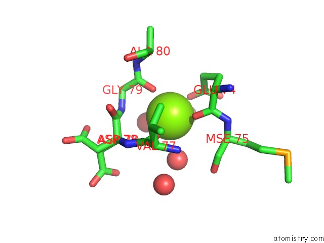
Mono view
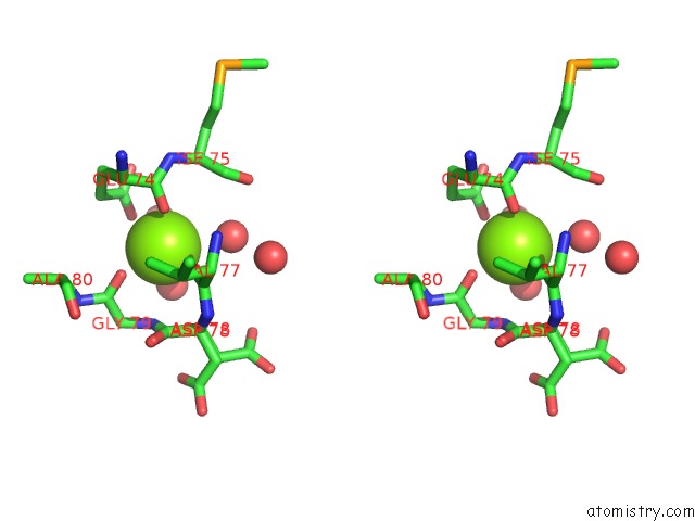
Stereo pair view
Mono view
Stereo pair view
A full contact list of Magnesium with other atoms in the Mg binding
site number 1 of Crystal Structure of Dsba-Like Thioredoxin Domain VF_A0457 From Vibrio Fischeri within 5.0Å range:
|
Reference:
Y.Kim,
A.Sather,
G.Shackelford,
A.Joachimiak.
Crystal Structure of Dsba-Like Thioredoxin Domain VF_A0457 From Vibrio Fischeri To Be Published.
Page generated: Wed Aug 14 13:46:08 2024
Last articles
Cl in 7U0NCl in 7U1D
Cl in 7U00
Cl in 7U0O
Cl in 7U0A
Cl in 7TZX
Cl in 7TZW
Cl in 7TZY
Cl in 7TXR
Cl in 7TZ6