Magnesium »
PDB 3fcw-3fpk »
3fkq »
Magnesium in PDB 3fkq: Crystal Structure of Ntrc-Like Two-Domain Protein (RER070207001320) From Eubacterium Rectale at 2.10 A Resolution
Protein crystallography data
The structure of Crystal Structure of Ntrc-Like Two-Domain Protein (RER070207001320) From Eubacterium Rectale at 2.10 A Resolution, PDB code: 3fkq
was solved by
Joint Center For Structural Genomics (Jcsg),
with X-Ray Crystallography technique. A brief refinement statistics is given in the table below:
| Resolution Low / High (Å) | 29.16 / 2.10 |
| Space group | C 1 2 1 |
| Cell size a, b, c (Å), α, β, γ (°) | 121.506, 79.495, 55.022, 90.00, 96.16, 90.00 |
| R / Rfree (%) | 19 / 21.8 |
Other elements in 3fkq:
The structure of Crystal Structure of Ntrc-Like Two-Domain Protein (RER070207001320) From Eubacterium Rectale at 2.10 A Resolution also contains other interesting chemical elements:
| Chlorine | (Cl) | 1 atom |
Magnesium Binding Sites:
The binding sites of Magnesium atom in the Crystal Structure of Ntrc-Like Two-Domain Protein (RER070207001320) From Eubacterium Rectale at 2.10 A Resolution
(pdb code 3fkq). This binding sites where shown within
5.0 Angstroms radius around Magnesium atom.
In total only one binding site of Magnesium was determined in the Crystal Structure of Ntrc-Like Two-Domain Protein (RER070207001320) From Eubacterium Rectale at 2.10 A Resolution, PDB code: 3fkq:
In total only one binding site of Magnesium was determined in the Crystal Structure of Ntrc-Like Two-Domain Protein (RER070207001320) From Eubacterium Rectale at 2.10 A Resolution, PDB code: 3fkq:
Magnesium binding site 1 out of 1 in 3fkq
Go back to
Magnesium binding site 1 out
of 1 in the Crystal Structure of Ntrc-Like Two-Domain Protein (RER070207001320) From Eubacterium Rectale at 2.10 A Resolution
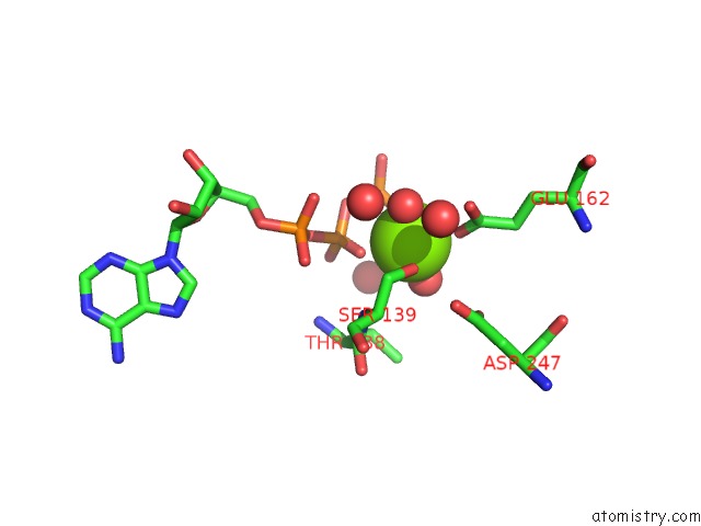
Mono view
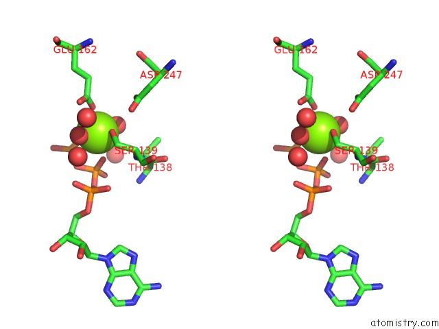
Stereo pair view
Mono view
Stereo pair view
A full contact list of Magnesium with other atoms in the Mg binding
site number 1 of Crystal Structure of Ntrc-Like Two-Domain Protein (RER070207001320) From Eubacterium Rectale at 2.10 A Resolution within 5.0Å range:
|
Reference:
Q.Xu,
B.Christen,
H.J.Chiu,
L.Jaroszewski,
H.E.Klock,
M.W.Knuth,
M.D.Miller,
M.A.Elsliger,
A.M.Deacon,
A.Godzik,
S.A.Lesley,
D.H.Figurski,
L.Shapiro,
I.A.Wilson.
Structure of the Pilus Assembly Protein Tadz From Eubacterium Rectale: Implications For Polar Localization. Mol.Microbiol. V. 83 712 2012.
ISSN: ISSN 0950-382X
PubMed: 22211578
DOI: 10.1111/J.1365-2958.2011.07954.X
Page generated: Wed Aug 14 13:49:15 2024
ISSN: ISSN 0950-382X
PubMed: 22211578
DOI: 10.1111/J.1365-2958.2011.07954.X
Last articles
Zn in 9MJ5Zn in 9HNW
Zn in 9G0L
Zn in 9FNE
Zn in 9DZN
Zn in 9E0I
Zn in 9D32
Zn in 9DAK
Zn in 8ZXC
Zn in 8ZUF