Magnesium »
PDB 3fps-3fzi »
3fs0 »
Magnesium in PDB 3fs0: Class II Ligase Ribozyme Product-Template Duplex, Structure 2
Protein crystallography data
The structure of Class II Ligase Ribozyme Product-Template Duplex, Structure 2, PDB code: 3fs0
was solved by
J.N.Pitt,
A.R.Ferre-D'amare,
with X-Ray Crystallography technique. A brief refinement statistics is given in the table below:
| Resolution Low / High (Å) | 23.85 / 2.30 |
| Space group | P 31 |
| Cell size a, b, c (Å), α, β, γ (°) | 30.040, 30.040, 59.666, 90.00, 90.00, 120.00 |
| R / Rfree (%) | 20.8 / 23.4 |
Magnesium Binding Sites:
The binding sites of Magnesium atom in the Class II Ligase Ribozyme Product-Template Duplex, Structure 2
(pdb code 3fs0). This binding sites where shown within
5.0 Angstroms radius around Magnesium atom.
In total 5 binding sites of Magnesium where determined in the Class II Ligase Ribozyme Product-Template Duplex, Structure 2, PDB code: 3fs0:
Jump to Magnesium binding site number: 1; 2; 3; 4; 5;
In total 5 binding sites of Magnesium where determined in the Class II Ligase Ribozyme Product-Template Duplex, Structure 2, PDB code: 3fs0:
Jump to Magnesium binding site number: 1; 2; 3; 4; 5;
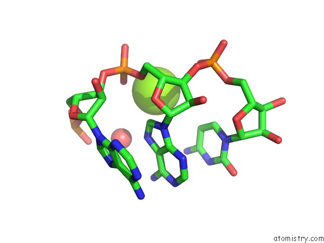
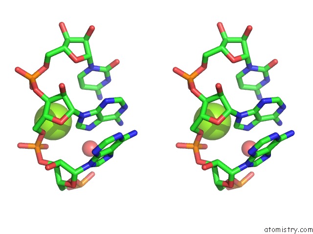
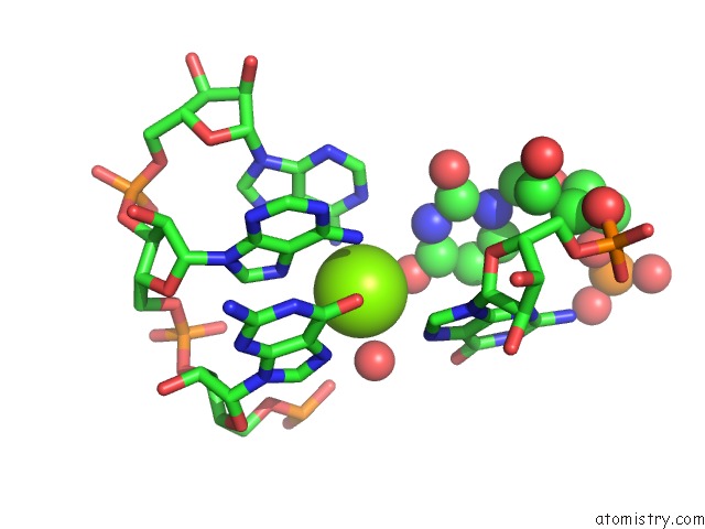
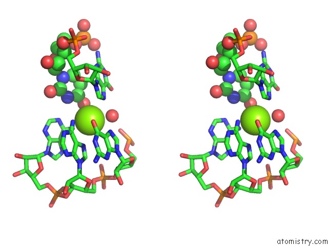
Magnesium binding site 1 out of 5 in 3fs0
Go back to
Magnesium binding site 1 out
of 5 in the Class II Ligase Ribozyme Product-Template Duplex, Structure 2
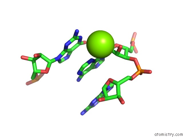
Mono view
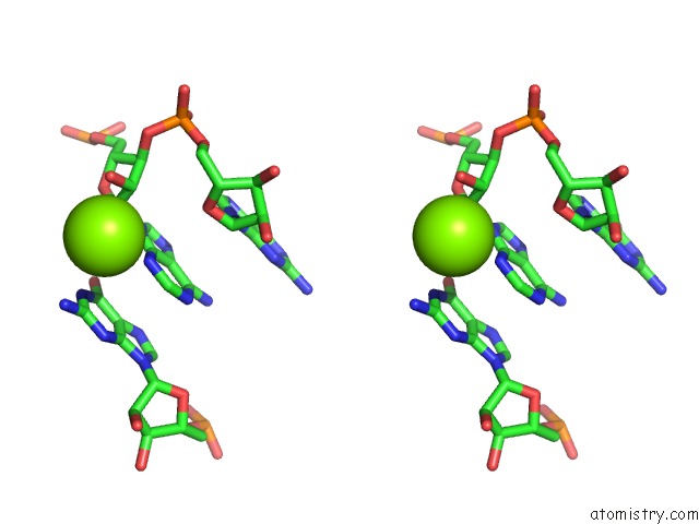
Stereo pair view
Mono view
Stereo pair view
A full contact list of Magnesium with other atoms in the Mg binding
site number 1 of Class II Ligase Ribozyme Product-Template Duplex, Structure 2 within 5.0Å range:
|
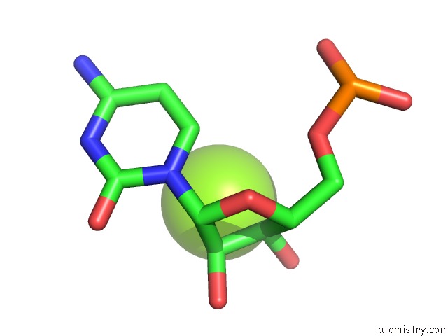
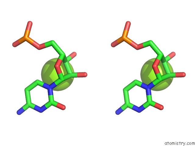
Magnesium binding site 2 out of 5 in 3fs0
Go back to
Magnesium binding site 2 out
of 5 in the Class II Ligase Ribozyme Product-Template Duplex, Structure 2
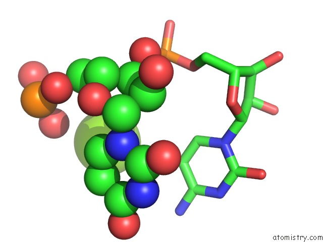
Mono view
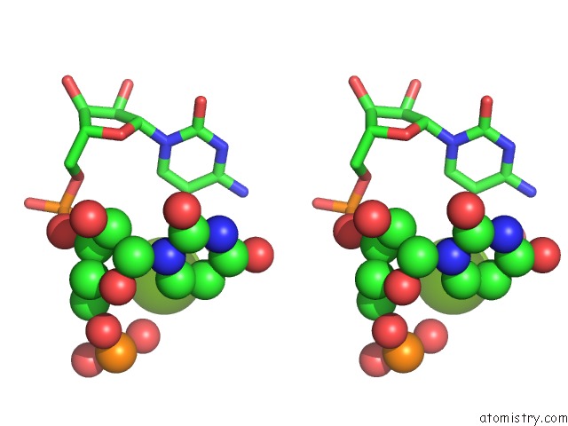
Stereo pair view
Mono view
Stereo pair view
A full contact list of Magnesium with other atoms in the Mg binding
site number 2 of Class II Ligase Ribozyme Product-Template Duplex, Structure 2 within 5.0Å range:
|
Magnesium binding site 3 out of 5 in 3fs0
Go back to
Magnesium binding site 3 out
of 5 in the Class II Ligase Ribozyme Product-Template Duplex, Structure 2
Mono view
Stereo pair view
Mono view
Stereo pair view
A full contact list of Magnesium with other atoms in the Mg binding
site number 3 of Class II Ligase Ribozyme Product-Template Duplex, Structure 2 within 5.0Å range:
|
Magnesium binding site 4 out of 5 in 3fs0
Go back to
Magnesium binding site 4 out
of 5 in the Class II Ligase Ribozyme Product-Template Duplex, Structure 2
Mono view
Stereo pair view
Mono view
Stereo pair view
A full contact list of Magnesium with other atoms in the Mg binding
site number 4 of Class II Ligase Ribozyme Product-Template Duplex, Structure 2 within 5.0Å range:
|
Magnesium binding site 5 out of 5 in 3fs0
Go back to
Magnesium binding site 5 out
of 5 in the Class II Ligase Ribozyme Product-Template Duplex, Structure 2
Mono view
Stereo pair view
Mono view
Stereo pair view
A full contact list of Magnesium with other atoms in the Mg binding
site number 5 of Class II Ligase Ribozyme Product-Template Duplex, Structure 2 within 5.0Å range:
|
Reference:
J.N.Pitt,
A.R.Ferre-D'amare.
Structure-Guided Engineering of the Regioselectivity of Rna Ligase Ribozymes. J.Am.Chem.Soc. V. 131 3532 2009.
ISSN: ISSN 0002-7863
PubMed: 19220054
DOI: 10.1021/JA8067325
Page generated: Wed Aug 14 13:52:50 2024
ISSN: ISSN 0002-7863
PubMed: 19220054
DOI: 10.1021/JA8067325
Last articles
Fe in 2YXOFe in 2YRS
Fe in 2YXC
Fe in 2YNM
Fe in 2YVJ
Fe in 2YP1
Fe in 2YU2
Fe in 2YU1
Fe in 2YQB
Fe in 2YOO