Magnesium »
PDB 3fps-3fzi »
3fwz »
Magnesium in PDB 3fwz: Crystal Structure of Trka-N Domain of Inner Membrane Protein Ybal From Escherichia Coli
Protein crystallography data
The structure of Crystal Structure of Trka-N Domain of Inner Membrane Protein Ybal From Escherichia Coli, PDB code: 3fwz
was solved by
C.Chang,
L.Bigelow,
K.Buck,
A.Joachimiak,
Midwest Center For Structuralgenomics (Mcsg),
with X-Ray Crystallography technique. A brief refinement statistics is given in the table below:
| Resolution Low / High (Å) | 50.00 / 1.79 |
| Space group | P 1 21 1 |
| Cell size a, b, c (Å), α, β, γ (°) | 34.801, 53.071, 72.537, 90.00, 97.82, 90.00 |
| R / Rfree (%) | 16.8 / 20.2 |
Magnesium Binding Sites:
The binding sites of Magnesium atom in the Crystal Structure of Trka-N Domain of Inner Membrane Protein Ybal From Escherichia Coli
(pdb code 3fwz). This binding sites where shown within
5.0 Angstroms radius around Magnesium atom.
In total only one binding site of Magnesium was determined in the Crystal Structure of Trka-N Domain of Inner Membrane Protein Ybal From Escherichia Coli, PDB code: 3fwz:
In total only one binding site of Magnesium was determined in the Crystal Structure of Trka-N Domain of Inner Membrane Protein Ybal From Escherichia Coli, PDB code: 3fwz:
Magnesium binding site 1 out of 1 in 3fwz
Go back to
Magnesium binding site 1 out
of 1 in the Crystal Structure of Trka-N Domain of Inner Membrane Protein Ybal From Escherichia Coli
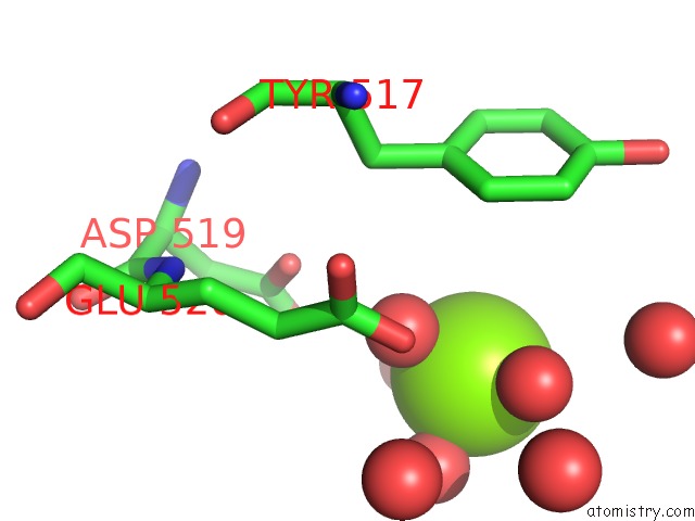
Mono view
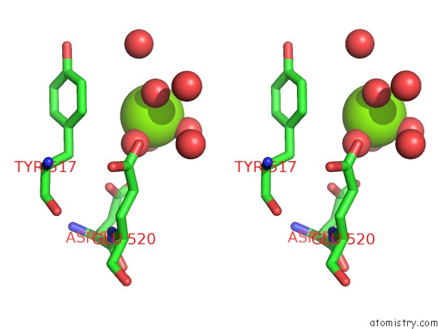
Stereo pair view
Mono view
Stereo pair view
A full contact list of Magnesium with other atoms in the Mg binding
site number 1 of Crystal Structure of Trka-N Domain of Inner Membrane Protein Ybal From Escherichia Coli within 5.0Å range:
|
Reference:
C.Chang,
L.Bigelow,
K.Buck,
A.Joachimiak.
Crystal Structure of Trka-N Domain of Inner Membrane Protein Ybal From Escherichia Coli To Be Published.
Page generated: Wed Aug 14 13:57:54 2024
Last articles
Fe in 2YXOFe in 2YRS
Fe in 2YXC
Fe in 2YNM
Fe in 2YVJ
Fe in 2YP1
Fe in 2YU2
Fe in 2YU1
Fe in 2YQB
Fe in 2YOO