Magnesium »
PDB 3fzn-3g8c »
3g5s »
Magnesium in PDB 3g5s: Crystal Structure of Thermus Thermophilus Trmfo in Complex with Glutathione
Enzymatic activity of Crystal Structure of Thermus Thermophilus Trmfo in Complex with Glutathione
All present enzymatic activity of Crystal Structure of Thermus Thermophilus Trmfo in Complex with Glutathione:
2.1.1.74;
2.1.1.74;
Protein crystallography data
The structure of Crystal Structure of Thermus Thermophilus Trmfo in Complex with Glutathione, PDB code: 3g5s
was solved by
H.Nishimasu,
R.Ishitani,
H.Hori,
O.Nureki,
with X-Ray Crystallography technique. A brief refinement statistics is given in the table below:
| Resolution Low / High (Å) | 43.81 / 1.05 |
| Space group | P 21 21 21 |
| Cell size a, b, c (Å), α, β, γ (°) | 48.257, 92.226, 104.501, 90.00, 90.00, 90.00 |
| R / Rfree (%) | 16.2 / 17.8 |
Magnesium Binding Sites:
The binding sites of Magnesium atom in the Crystal Structure of Thermus Thermophilus Trmfo in Complex with Glutathione
(pdb code 3g5s). This binding sites where shown within
5.0 Angstroms radius around Magnesium atom.
In total only one binding site of Magnesium was determined in the Crystal Structure of Thermus Thermophilus Trmfo in Complex with Glutathione, PDB code: 3g5s:
In total only one binding site of Magnesium was determined in the Crystal Structure of Thermus Thermophilus Trmfo in Complex with Glutathione, PDB code: 3g5s:
Magnesium binding site 1 out of 1 in 3g5s
Go back to
Magnesium binding site 1 out
of 1 in the Crystal Structure of Thermus Thermophilus Trmfo in Complex with Glutathione
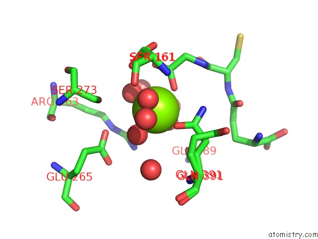
Mono view
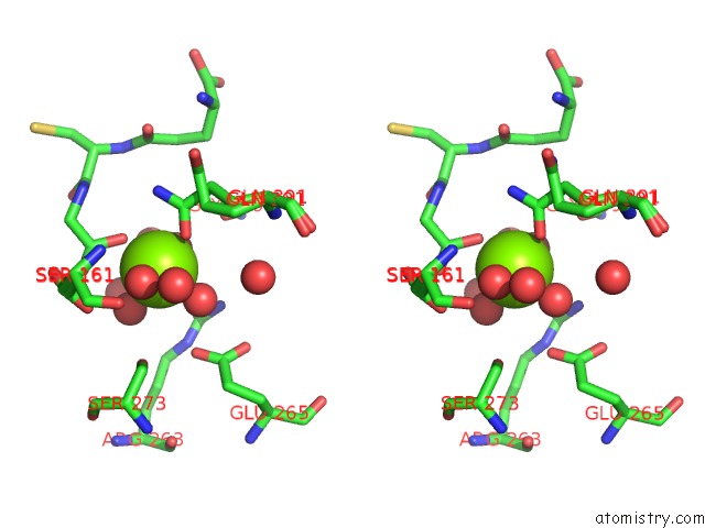
Stereo pair view
Mono view
Stereo pair view
A full contact list of Magnesium with other atoms in the Mg binding
site number 1 of Crystal Structure of Thermus Thermophilus Trmfo in Complex with Glutathione within 5.0Å range:
|
Reference:
H.Nishimasu,
R.Ishitani,
K.Yamashita,
C.Iwashita,
A.Hirata,
H.Hori,
O.Nureki.
Atomic Structure of A Folate/Fad-Dependent Trna T54 Methyltransferase Proc.Natl.Acad.Sci.Usa V. 106 8180 2009.
ISSN: ISSN 0027-8424
PubMed: 19416846
DOI: 10.1073/PNAS.0901330106
Page generated: Sun Aug 10 21:13:53 2025
ISSN: ISSN 0027-8424
PubMed: 19416846
DOI: 10.1073/PNAS.0901330106
Last articles
Mg in 6DN1Mg in 6DMV
Mg in 6DMC
Mg in 6DMD
Mg in 6DLS
Mg in 6DLT
Mg in 6DLU
Mg in 6DLR
Mg in 6DKO
Mg in 6DL9