Magnesium »
PDB 3fzn-3g8c »
3g8c »
Magnesium in PDB 3g8c: Crystal Structure of Biotin Carboxylase in Complex with Biotin, Bicarbonate, Adp and Mg Ion
Enzymatic activity of Crystal Structure of Biotin Carboxylase in Complex with Biotin, Bicarbonate, Adp and Mg Ion
All present enzymatic activity of Crystal Structure of Biotin Carboxylase in Complex with Biotin, Bicarbonate, Adp and Mg Ion:
6.3.4.14; 6.4.1.2;
6.3.4.14; 6.4.1.2;
Protein crystallography data
The structure of Crystal Structure of Biotin Carboxylase in Complex with Biotin, Bicarbonate, Adp and Mg Ion, PDB code: 3g8c
was solved by
C.Y.Chou,
L.P.Yu,
L.Tong,
with X-Ray Crystallography technique. A brief refinement statistics is given in the table below:
| Resolution Low / High (Å) | 30.00 / 2.00 |
| Space group | P 21 21 21 |
| Cell size a, b, c (Å), α, β, γ (°) | 83.317, 106.166, 121.486, 90.00, 90.00, 90.00 |
| R / Rfree (%) | 18.2 / 21.6 |
Magnesium Binding Sites:
The binding sites of Magnesium atom in the Crystal Structure of Biotin Carboxylase in Complex with Biotin, Bicarbonate, Adp and Mg Ion
(pdb code 3g8c). This binding sites where shown within
5.0 Angstroms radius around Magnesium atom.
In total 2 binding sites of Magnesium where determined in the Crystal Structure of Biotin Carboxylase in Complex with Biotin, Bicarbonate, Adp and Mg Ion, PDB code: 3g8c:
Jump to Magnesium binding site number: 1; 2;
In total 2 binding sites of Magnesium where determined in the Crystal Structure of Biotin Carboxylase in Complex with Biotin, Bicarbonate, Adp and Mg Ion, PDB code: 3g8c:
Jump to Magnesium binding site number: 1; 2;
Magnesium binding site 1 out of 2 in 3g8c
Go back to
Magnesium binding site 1 out
of 2 in the Crystal Structure of Biotin Carboxylase in Complex with Biotin, Bicarbonate, Adp and Mg Ion
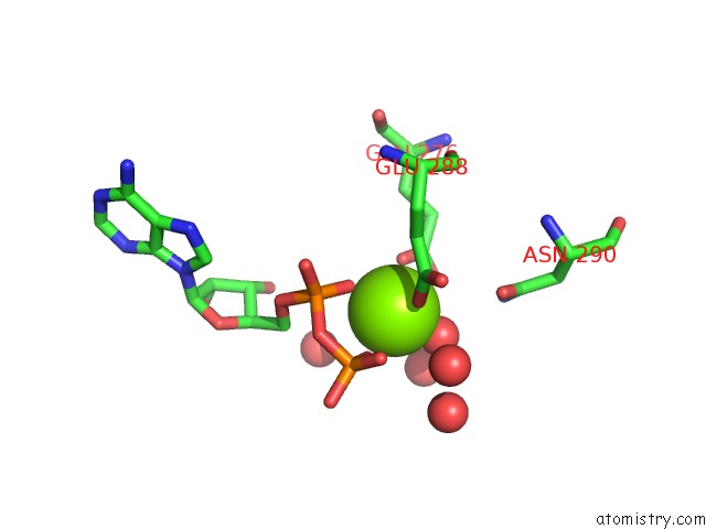
Mono view
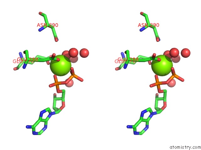
Stereo pair view
Mono view
Stereo pair view
A full contact list of Magnesium with other atoms in the Mg binding
site number 1 of Crystal Structure of Biotin Carboxylase in Complex with Biotin, Bicarbonate, Adp and Mg Ion within 5.0Å range:
|
Magnesium binding site 2 out of 2 in 3g8c
Go back to
Magnesium binding site 2 out
of 2 in the Crystal Structure of Biotin Carboxylase in Complex with Biotin, Bicarbonate, Adp and Mg Ion
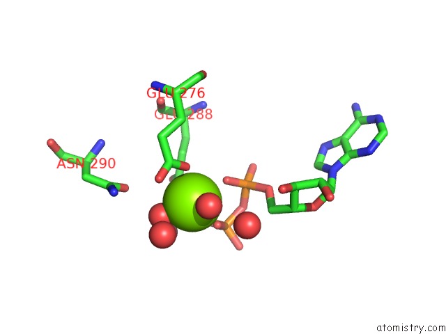
Mono view
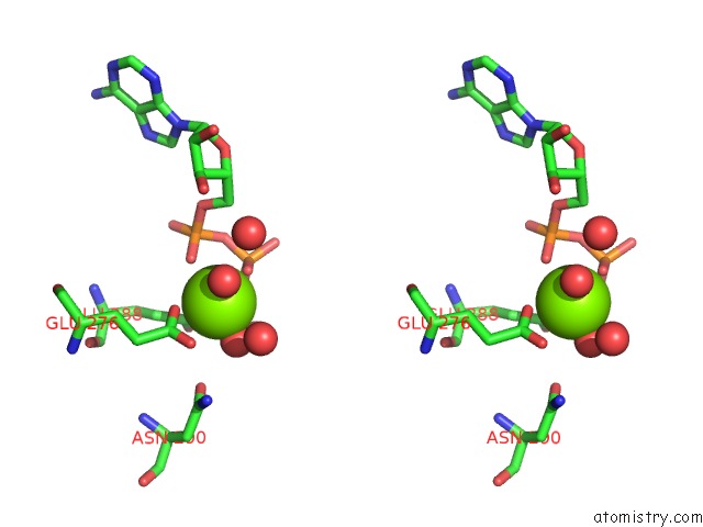
Stereo pair view
Mono view
Stereo pair view
A full contact list of Magnesium with other atoms in the Mg binding
site number 2 of Crystal Structure of Biotin Carboxylase in Complex with Biotin, Bicarbonate, Adp and Mg Ion within 5.0Å range:
|
Reference:
C.Y.Chou,
L.P.Yu,
L.Tong.
Crystal Structure of Biotin Carboxylase in Complex with Substrates and Implications For Its Catalytic Mechanism. J.Biol.Chem. V. 284 11690 2009.
ISSN: ISSN 0021-9258
PubMed: 19213731
DOI: 10.1074/JBC.M805783200
Page generated: Sun Aug 10 21:34:23 2025
ISSN: ISSN 0021-9258
PubMed: 19213731
DOI: 10.1074/JBC.M805783200
Last articles
Mg in 6TRAMg in 6TR4
Mg in 6TR3
Mg in 6TMF
Mg in 6TQO
Mg in 6TQN
Mg in 6TQF
Mg in 6TQE
Mg in 6TQB
Mg in 6TQA