Magnesium »
PDB 3g8d-3gn6 »
3g8t »
Magnesium in PDB 3g8t: Crystal Structure of the G33A Mutant Bacillus Anthracis Glms Ribozyme Bound to GLCN6P
Protein crystallography data
The structure of Crystal Structure of the G33A Mutant Bacillus Anthracis Glms Ribozyme Bound to GLCN6P, PDB code: 3g8t
was solved by
S.A.Strobel,
J.C.Cochrane,
S.V.Lipchock,
K.D.Smith,
with X-Ray Crystallography technique. A brief refinement statistics is given in the table below:
| Resolution Low / High (Å) | 28.23 / 3.00 |
| Space group | P 1 21 1 |
| Cell size a, b, c (Å), α, β, γ (°) | 48.389, 232.671, 106.576, 90.00, 92.21, 90.00 |
| R / Rfree (%) | 25 / 31.8 |
Magnesium Binding Sites:
The binding sites of Magnesium atom in the Crystal Structure of the G33A Mutant Bacillus Anthracis Glms Ribozyme Bound to GLCN6P
(pdb code 3g8t). This binding sites where shown within
5.0 Angstroms radius around Magnesium atom.
In total 10 binding sites of Magnesium where determined in the Crystal Structure of the G33A Mutant Bacillus Anthracis Glms Ribozyme Bound to GLCN6P, PDB code: 3g8t:
Jump to Magnesium binding site number: 1; 2; 3; 4; 5; 6; 7; 8; 9; 10;
In total 10 binding sites of Magnesium where determined in the Crystal Structure of the G33A Mutant Bacillus Anthracis Glms Ribozyme Bound to GLCN6P, PDB code: 3g8t:
Jump to Magnesium binding site number: 1; 2; 3; 4; 5; 6; 7; 8; 9; 10;
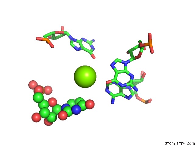
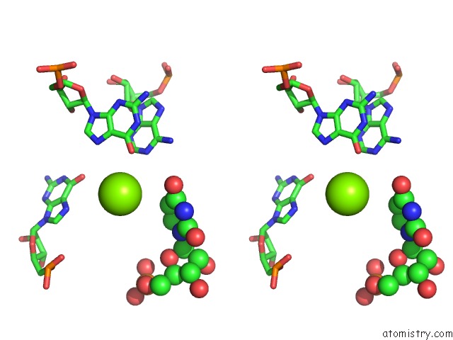
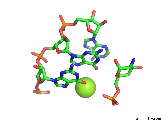
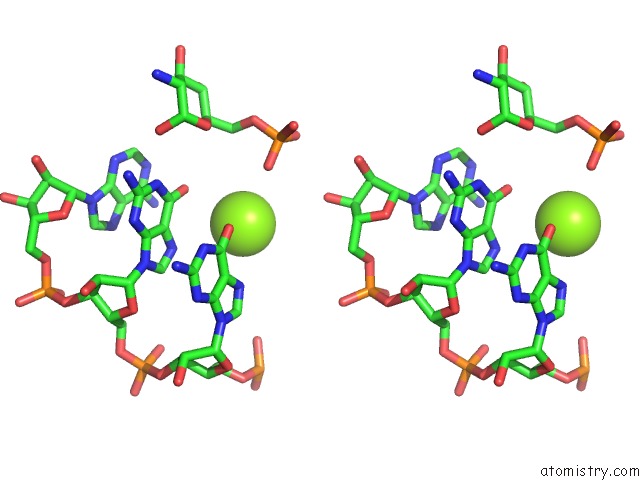
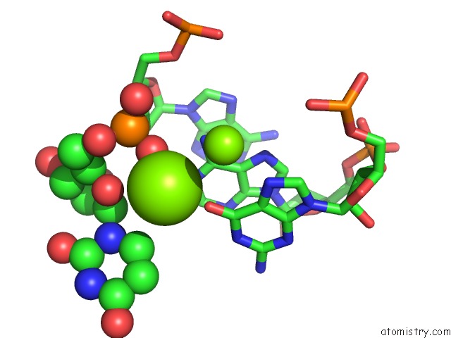
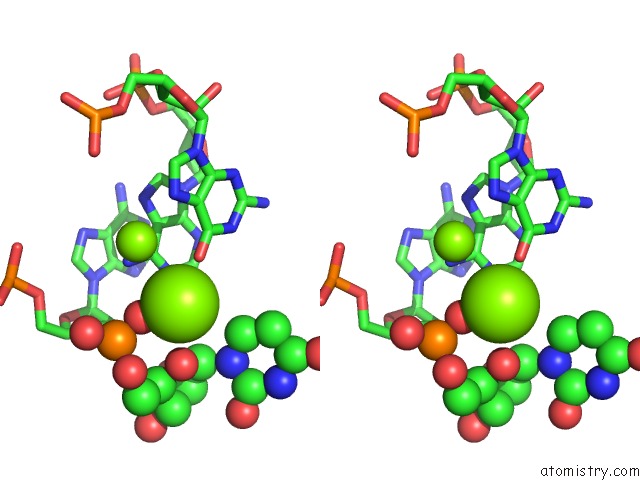
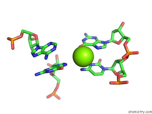
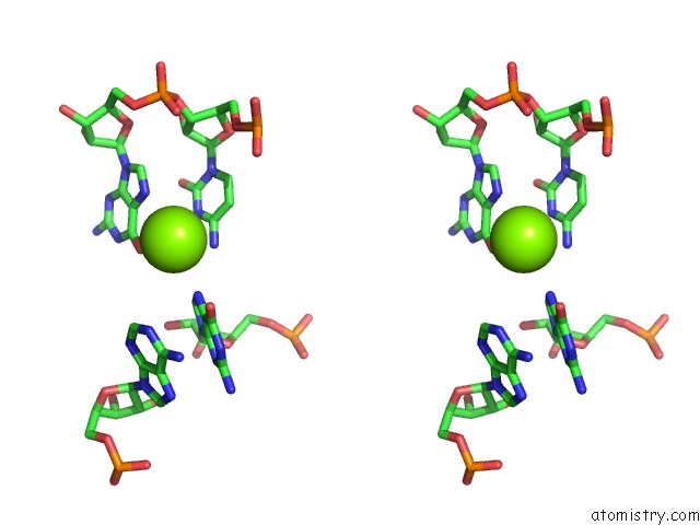
Magnesium binding site 1 out of 10 in 3g8t
Go back to
Magnesium binding site 1 out
of 10 in the Crystal Structure of the G33A Mutant Bacillus Anthracis Glms Ribozyme Bound to GLCN6P
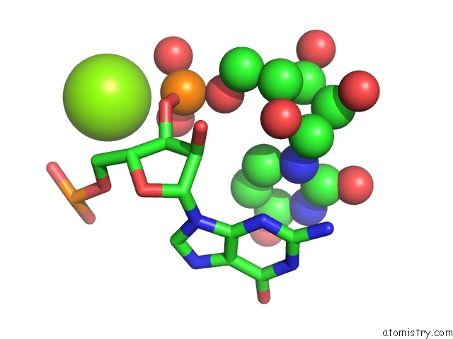
Mono view
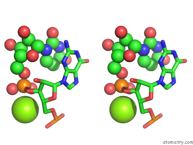
Stereo pair view
Mono view
Stereo pair view
A full contact list of Magnesium with other atoms in the Mg binding
site number 1 of Crystal Structure of the G33A Mutant Bacillus Anthracis Glms Ribozyme Bound to GLCN6P within 5.0Å range:
|
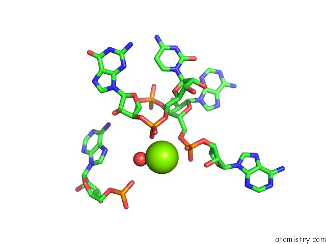
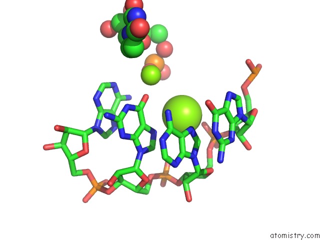
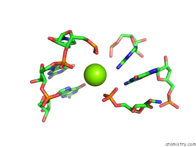
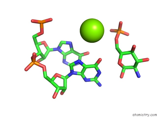
Magnesium binding site 2 out of 10 in 3g8t
Go back to
Magnesium binding site 2 out
of 10 in the Crystal Structure of the G33A Mutant Bacillus Anthracis Glms Ribozyme Bound to GLCN6P
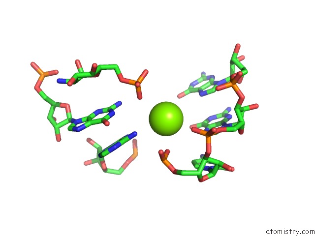
Mono view
Stereo pair view
Mono view
Stereo pair view
A full contact list of Magnesium with other atoms in the Mg binding
site number 2 of Crystal Structure of the G33A Mutant Bacillus Anthracis Glms Ribozyme Bound to GLCN6P within 5.0Å range:
|
Magnesium binding site 3 out of 10 in 3g8t
Go back to
Magnesium binding site 3 out
of 10 in the Crystal Structure of the G33A Mutant Bacillus Anthracis Glms Ribozyme Bound to GLCN6P
Mono view
Stereo pair view
Mono view
Stereo pair view
A full contact list of Magnesium with other atoms in the Mg binding
site number 3 of Crystal Structure of the G33A Mutant Bacillus Anthracis Glms Ribozyme Bound to GLCN6P within 5.0Å range:
|
Magnesium binding site 4 out of 10 in 3g8t
Go back to
Magnesium binding site 4 out
of 10 in the Crystal Structure of the G33A Mutant Bacillus Anthracis Glms Ribozyme Bound to GLCN6P
Mono view
Stereo pair view
Mono view
Stereo pair view
A full contact list of Magnesium with other atoms in the Mg binding
site number 4 of Crystal Structure of the G33A Mutant Bacillus Anthracis Glms Ribozyme Bound to GLCN6P within 5.0Å range:
|
Magnesium binding site 5 out of 10 in 3g8t
Go back to
Magnesium binding site 5 out
of 10 in the Crystal Structure of the G33A Mutant Bacillus Anthracis Glms Ribozyme Bound to GLCN6P
Mono view
Stereo pair view
Mono view
Stereo pair view
A full contact list of Magnesium with other atoms in the Mg binding
site number 5 of Crystal Structure of the G33A Mutant Bacillus Anthracis Glms Ribozyme Bound to GLCN6P within 5.0Å range:
|
Magnesium binding site 6 out of 10 in 3g8t
Go back to
Magnesium binding site 6 out
of 10 in the Crystal Structure of the G33A Mutant Bacillus Anthracis Glms Ribozyme Bound to GLCN6P
Mono view
Stereo pair view
Mono view
Stereo pair view
A full contact list of Magnesium with other atoms in the Mg binding
site number 6 of Crystal Structure of the G33A Mutant Bacillus Anthracis Glms Ribozyme Bound to GLCN6P within 5.0Å range:
|
Magnesium binding site 7 out of 10 in 3g8t
Go back to
Magnesium binding site 7 out
of 10 in the Crystal Structure of the G33A Mutant Bacillus Anthracis Glms Ribozyme Bound to GLCN6P
Mono view
Stereo pair view
Mono view
Stereo pair view
A full contact list of Magnesium with other atoms in the Mg binding
site number 7 of Crystal Structure of the G33A Mutant Bacillus Anthracis Glms Ribozyme Bound to GLCN6P within 5.0Å range:
|
Magnesium binding site 8 out of 10 in 3g8t
Go back to
Magnesium binding site 8 out
of 10 in the Crystal Structure of the G33A Mutant Bacillus Anthracis Glms Ribozyme Bound to GLCN6P
Mono view
Stereo pair view
Mono view
Stereo pair view
A full contact list of Magnesium with other atoms in the Mg binding
site number 8 of Crystal Structure of the G33A Mutant Bacillus Anthracis Glms Ribozyme Bound to GLCN6P within 5.0Å range:
|
Magnesium binding site 9 out of 10 in 3g8t
Go back to
Magnesium binding site 9 out
of 10 in the Crystal Structure of the G33A Mutant Bacillus Anthracis Glms Ribozyme Bound to GLCN6P
Mono view
Stereo pair view
Mono view
Stereo pair view
A full contact list of Magnesium with other atoms in the Mg binding
site number 9 of Crystal Structure of the G33A Mutant Bacillus Anthracis Glms Ribozyme Bound to GLCN6P within 5.0Å range:
|
Magnesium binding site 10 out of 10 in 3g8t
Go back to
Magnesium binding site 10 out
of 10 in the Crystal Structure of the G33A Mutant Bacillus Anthracis Glms Ribozyme Bound to GLCN6P
Mono view
Stereo pair view
Mono view
Stereo pair view
A full contact list of Magnesium with other atoms in the Mg binding
site number 10 of Crystal Structure of the G33A Mutant Bacillus Anthracis Glms Ribozyme Bound to GLCN6P within 5.0Å range:
|
Reference:
J.C.Cochrane,
S.V.Lipchock,
K.D.Smith,
S.A.Strobel.
Structural and Chemical Basis For Glucosamine 6-Phosphate Binding and Activation of the Glms Ribozyme Biochemistry V. 48 3239 2009.
ISSN: ISSN 0006-2960
PubMed: 19228039
DOI: 10.1021/BI802069P
Page generated: Wed Aug 14 14:41:29 2024
ISSN: ISSN 0006-2960
PubMed: 19228039
DOI: 10.1021/BI802069P
Last articles
F in 4MOFF in 4LZZ
F in 4LY6
F in 4MM8
F in 4MIH
F in 4MM4
F in 4MK0
F in 4MIG
F in 4MH0
F in 4MDD