Magnesium »
PDB 3g8d-3gn6 »
3g91 »
Magnesium in PDB 3g91: 1.2 Angstrom Structure of the Exonuclease III Homologue MTH0212
Enzymatic activity of 1.2 Angstrom Structure of the Exonuclease III Homologue MTH0212
All present enzymatic activity of 1.2 Angstrom Structure of the Exonuclease III Homologue MTH0212:
3.1.11.2;
3.1.11.2;
Protein crystallography data
The structure of 1.2 Angstrom Structure of the Exonuclease III Homologue MTH0212, PDB code: 3g91
was solved by
K.Lakomek,
A.Dickmanns,
R.Ficner,
with X-Ray Crystallography technique. A brief refinement statistics is given in the table below:
| Resolution Low / High (Å) | 9.99 / 1.23 |
| Space group | P 1 21 1 |
| Cell size a, b, c (Å), α, β, γ (°) | 44.330, 72.110, 46.330, 90.00, 117.96, 90.00 |
| R / Rfree (%) | 12.2 / 17.2 |
Magnesium Binding Sites:
The binding sites of Magnesium atom in the 1.2 Angstrom Structure of the Exonuclease III Homologue MTH0212
(pdb code 3g91). This binding sites where shown within
5.0 Angstroms radius around Magnesium atom.
In total only one binding site of Magnesium was determined in the 1.2 Angstrom Structure of the Exonuclease III Homologue MTH0212, PDB code: 3g91:
In total only one binding site of Magnesium was determined in the 1.2 Angstrom Structure of the Exonuclease III Homologue MTH0212, PDB code: 3g91:
Magnesium binding site 1 out of 1 in 3g91
Go back to
Magnesium binding site 1 out
of 1 in the 1.2 Angstrom Structure of the Exonuclease III Homologue MTH0212
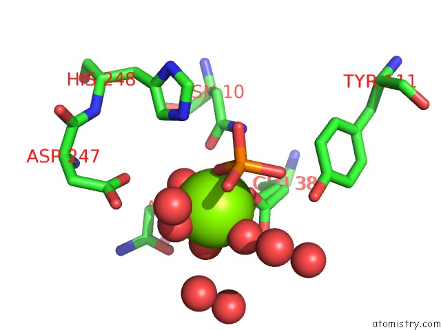
Mono view
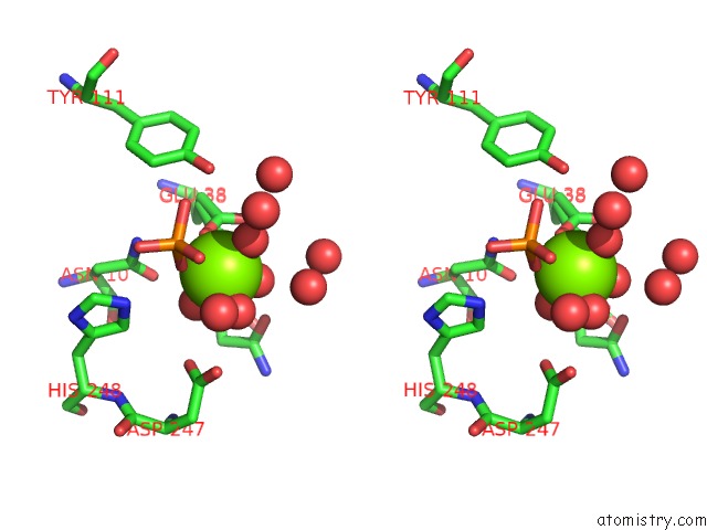
Stereo pair view
Mono view
Stereo pair view
A full contact list of Magnesium with other atoms in the Mg binding
site number 1 of 1.2 Angstrom Structure of the Exonuclease III Homologue MTH0212 within 5.0Å range:
|
Reference:
K.Lakomek,
A.Dickmanns,
E.Ciirdaeva,
L.Schomacher,
R.Ficner.
Crystal Structure Analysis of Dna Uridine Endonuclease MTH212 Bound to Dna J.Mol.Biol. V. 399 604 2010.
ISSN: ISSN 0022-2836
PubMed: 20434457
DOI: 10.1016/J.JMB.2010.04.044
Page generated: Wed Aug 14 14:41:28 2024
ISSN: ISSN 0022-2836
PubMed: 20434457
DOI: 10.1016/J.JMB.2010.04.044
Last articles
Cl in 8AK8Cl in 8AK7
Cl in 8AJP
Cl in 8AJ6
Cl in 8AIO
Cl in 8AI7
Cl in 8AHZ
Cl in 8AHQ
Cl in 8AHY
Cl in 8AHO