Magnesium »
PDB 3g8d-3gn6 »
3gah »
Magnesium in PDB 3gah: Structure of A F112H Variant Pduo-Type Atp:Corrinoid Adenosyltransferase From Lactobacillus Reuteri Complexed with Cobalamin and Atp
Enzymatic activity of Structure of A F112H Variant Pduo-Type Atp:Corrinoid Adenosyltransferase From Lactobacillus Reuteri Complexed with Cobalamin and Atp
All present enzymatic activity of Structure of A F112H Variant Pduo-Type Atp:Corrinoid Adenosyltransferase From Lactobacillus Reuteri Complexed with Cobalamin and Atp:
2.5.1.17;
2.5.1.17;
Protein crystallography data
The structure of Structure of A F112H Variant Pduo-Type Atp:Corrinoid Adenosyltransferase From Lactobacillus Reuteri Complexed with Cobalamin and Atp, PDB code: 3gah
was solved by
M.St Maurice,
P.E.Mera,
J.C.Escalante-Semerena,
I.Rayment,
with X-Ray Crystallography technique. A brief refinement statistics is given in the table below:
| Resolution Low / High (Å) | 30.00 / 1.17 |
| Space group | H 3 |
| Cell size a, b, c (Å), α, β, γ (°) | 80.814, 80.814, 89.492, 90.00, 90.00, 120.00 |
| R / Rfree (%) | 16 / 18.2 |
Other elements in 3gah:
The structure of Structure of A F112H Variant Pduo-Type Atp:Corrinoid Adenosyltransferase From Lactobacillus Reuteri Complexed with Cobalamin and Atp also contains other interesting chemical elements:
| Cobalt | (Co) | 2 atoms |
| Potassium | (K) | 1 atom |
Magnesium Binding Sites:
The binding sites of Magnesium atom in the Structure of A F112H Variant Pduo-Type Atp:Corrinoid Adenosyltransferase From Lactobacillus Reuteri Complexed with Cobalamin and Atp
(pdb code 3gah). This binding sites where shown within
5.0 Angstroms radius around Magnesium atom.
In total 2 binding sites of Magnesium where determined in the Structure of A F112H Variant Pduo-Type Atp:Corrinoid Adenosyltransferase From Lactobacillus Reuteri Complexed with Cobalamin and Atp, PDB code: 3gah:
Jump to Magnesium binding site number: 1; 2;
In total 2 binding sites of Magnesium where determined in the Structure of A F112H Variant Pduo-Type Atp:Corrinoid Adenosyltransferase From Lactobacillus Reuteri Complexed with Cobalamin and Atp, PDB code: 3gah:
Jump to Magnesium binding site number: 1; 2;
Magnesium binding site 1 out of 2 in 3gah
Go back to
Magnesium binding site 1 out
of 2 in the Structure of A F112H Variant Pduo-Type Atp:Corrinoid Adenosyltransferase From Lactobacillus Reuteri Complexed with Cobalamin and Atp
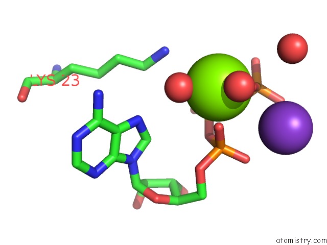
Mono view
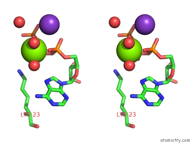
Stereo pair view
Mono view
Stereo pair view
A full contact list of Magnesium with other atoms in the Mg binding
site number 1 of Structure of A F112H Variant Pduo-Type Atp:Corrinoid Adenosyltransferase From Lactobacillus Reuteri Complexed with Cobalamin and Atp within 5.0Å range:
|
Magnesium binding site 2 out of 2 in 3gah
Go back to
Magnesium binding site 2 out
of 2 in the Structure of A F112H Variant Pduo-Type Atp:Corrinoid Adenosyltransferase From Lactobacillus Reuteri Complexed with Cobalamin and Atp
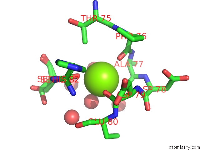
Mono view
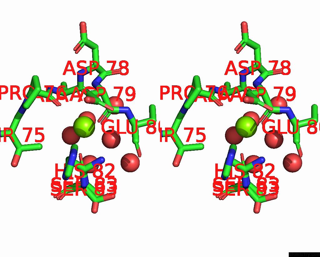
Stereo pair view
Mono view
Stereo pair view
A full contact list of Magnesium with other atoms in the Mg binding
site number 2 of Structure of A F112H Variant Pduo-Type Atp:Corrinoid Adenosyltransferase From Lactobacillus Reuteri Complexed with Cobalamin and Atp within 5.0Å range:
|
Reference:
P.E.Mera,
M.St Maurice,
I.Rayment,
J.C.Escalante-Semerena.
Residue PHE112 of the Human-Type Corrinoid Adenosyltransferase (Pduo) Enzyme of Lactobacillus Reuteri Is Critical to the Formation of the Four-Coordinate Co(II) Corrinoid Substrate and to the Activity of the Enzyme. Biochemistry V. 48 3138 2009.
ISSN: ISSN 0006-2960
PubMed: 19236001
DOI: 10.1021/BI9000134
Page generated: Wed Aug 14 14:41:55 2024
ISSN: ISSN 0006-2960
PubMed: 19236001
DOI: 10.1021/BI9000134
Last articles
Zn in 9MJ5Zn in 9HNW
Zn in 9G0L
Zn in 9FNE
Zn in 9DZN
Zn in 9E0I
Zn in 9D32
Zn in 9DAK
Zn in 8ZXC
Zn in 8ZUF