Magnesium »
PDB 3g8d-3gn6 »
3gli »
Magnesium in PDB 3gli: Crystal Structure of the E. Coli Clamp Loader Bound to Primer-Template Dna and Psi Peptide
Enzymatic activity of Crystal Structure of the E. Coli Clamp Loader Bound to Primer-Template Dna and Psi Peptide
All present enzymatic activity of Crystal Structure of the E. Coli Clamp Loader Bound to Primer-Template Dna and Psi Peptide:
2.7.7.7;
2.7.7.7;
Protein crystallography data
The structure of Crystal Structure of the E. Coli Clamp Loader Bound to Primer-Template Dna and Psi Peptide, PDB code: 3gli
was solved by
K.R.Simonetta,
A.J.Cantor,
J.Kuriyan,
with X-Ray Crystallography technique. A brief refinement statistics is given in the table below:
| Resolution Low / High (Å) | 73.03 / 3.50 |
| Space group | P 21 21 21 |
| Cell size a, b, c (Å), α, β, γ (°) | 98.690, 217.180, 275.320, 90.00, 90.00, 90.00 |
| R / Rfree (%) | 22.2 / 25.7 |
Other elements in 3gli:
The structure of Crystal Structure of the E. Coli Clamp Loader Bound to Primer-Template Dna and Psi Peptide also contains other interesting chemical elements:
| Fluorine | (F) | 18 atoms |
| Zinc | (Zn) | 8 atoms |
Magnesium Binding Sites:
The binding sites of Magnesium atom in the Crystal Structure of the E. Coli Clamp Loader Bound to Primer-Template Dna and Psi Peptide
(pdb code 3gli). This binding sites where shown within
5.0 Angstroms radius around Magnesium atom.
In total 6 binding sites of Magnesium where determined in the Crystal Structure of the E. Coli Clamp Loader Bound to Primer-Template Dna and Psi Peptide, PDB code: 3gli:
Jump to Magnesium binding site number: 1; 2; 3; 4; 5; 6;
In total 6 binding sites of Magnesium where determined in the Crystal Structure of the E. Coli Clamp Loader Bound to Primer-Template Dna and Psi Peptide, PDB code: 3gli:
Jump to Magnesium binding site number: 1; 2; 3; 4; 5; 6;
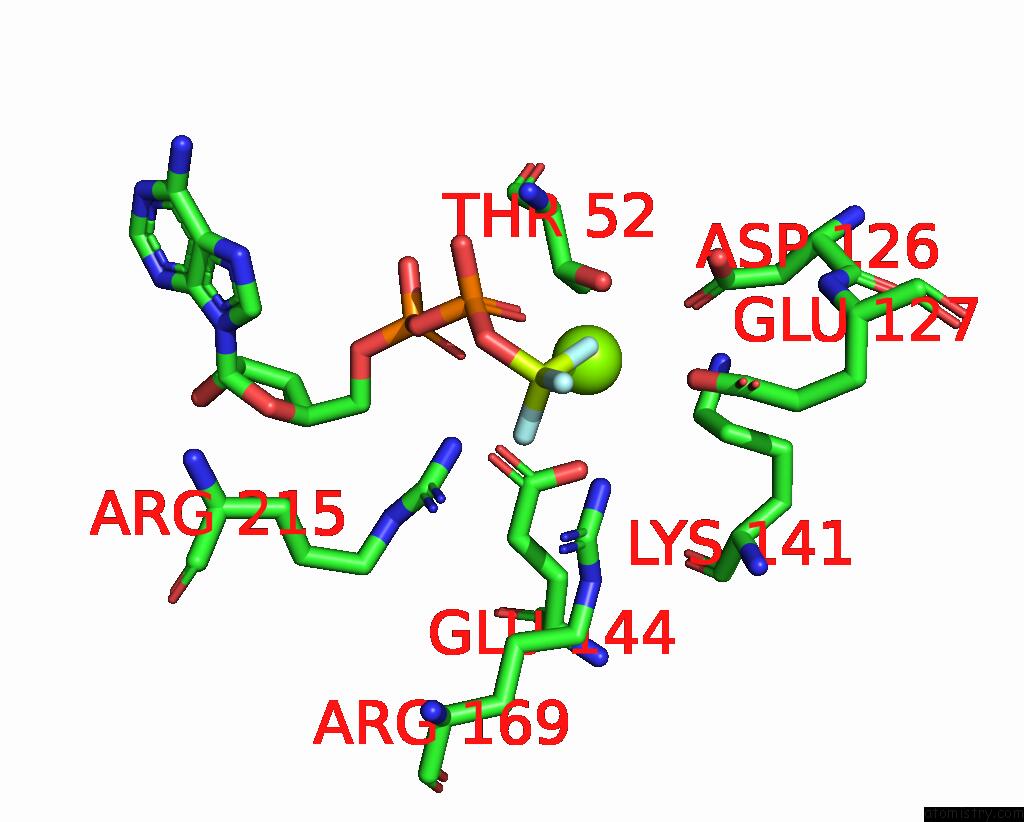
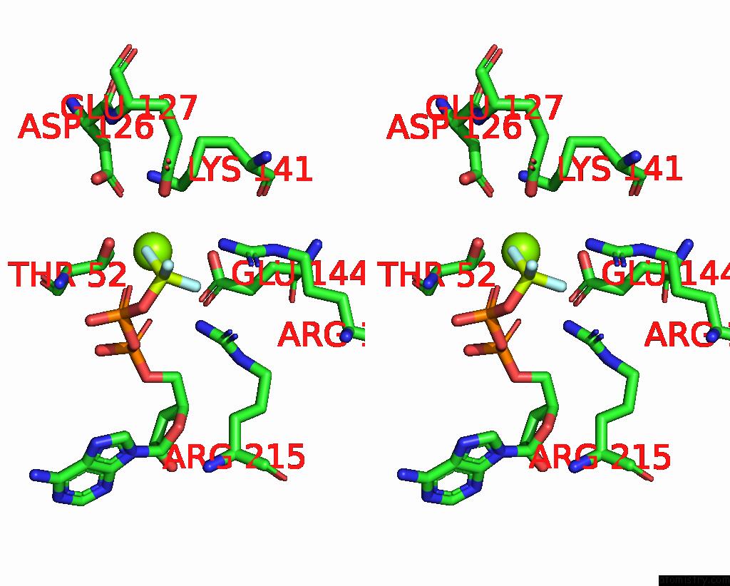
Magnesium binding site 1 out of 6 in 3gli
Go back to
Magnesium binding site 1 out
of 6 in the Crystal Structure of the E. Coli Clamp Loader Bound to Primer-Template Dna and Psi Peptide
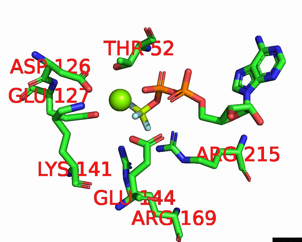
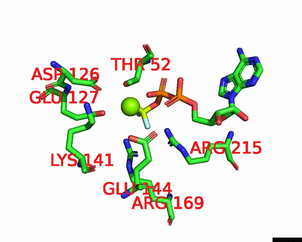
Mono view

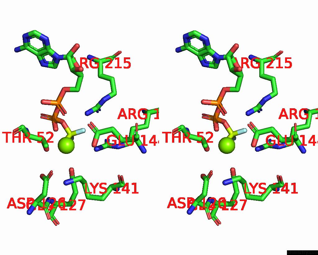
Stereo pair view
Mono view
Stereo pair view
A full contact list of Magnesium with other atoms in the Mg binding
site number 1 of Crystal Structure of the E. Coli Clamp Loader Bound to Primer-Template Dna and Psi Peptide within 5.0Å range:
|
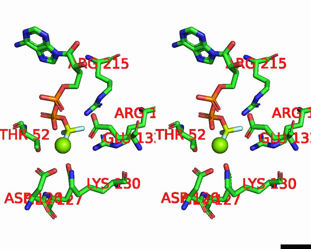
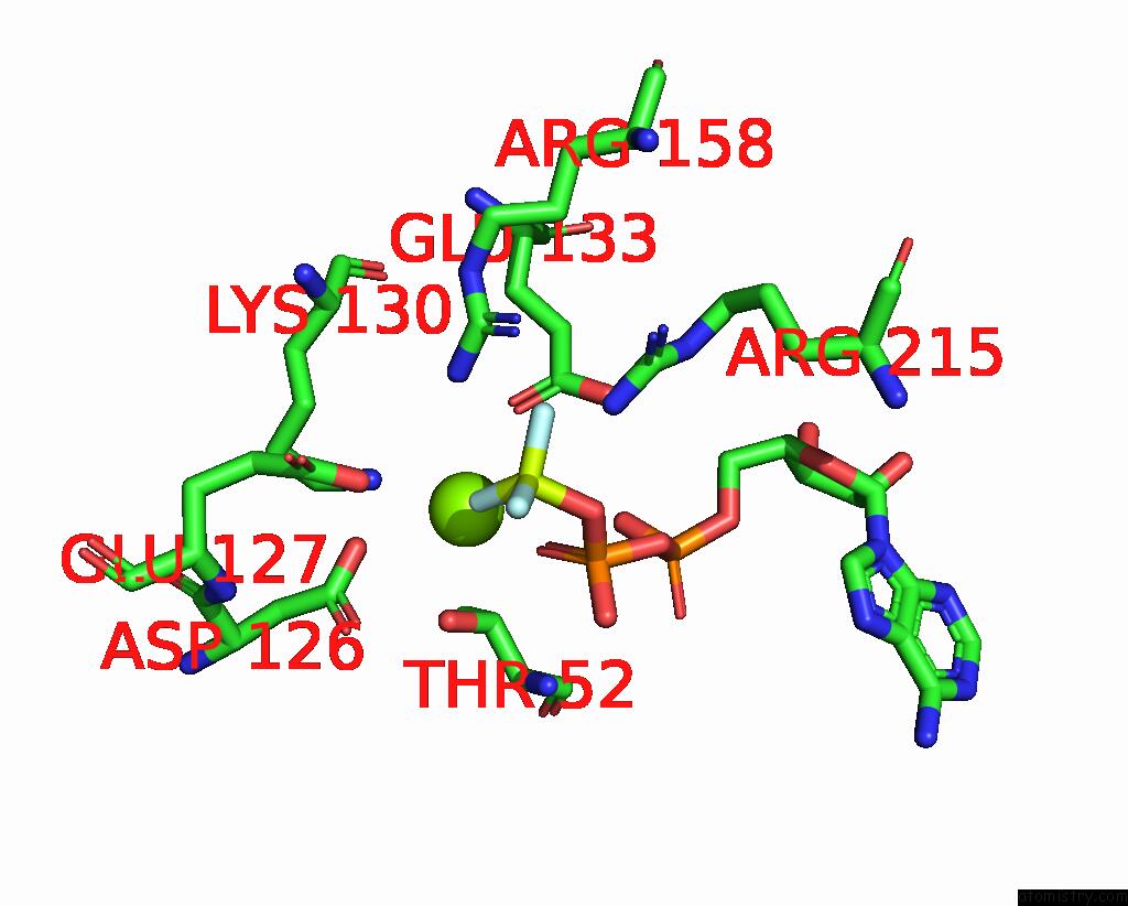
Magnesium binding site 2 out of 6 in 3gli
Go back to
Magnesium binding site 2 out
of 6 in the Crystal Structure of the E. Coli Clamp Loader Bound to Primer-Template Dna and Psi Peptide
Mono view
Stereo pair view
Mono view
Stereo pair view
A full contact list of Magnesium with other atoms in the Mg binding
site number 2 of Crystal Structure of the E. Coli Clamp Loader Bound to Primer-Template Dna and Psi Peptide within 5.0Å range:
|
Magnesium binding site 3 out of 6 in 3gli
Go back to
Magnesium binding site 3 out
of 6 in the Crystal Structure of the E. Coli Clamp Loader Bound to Primer-Template Dna and Psi Peptide

Mono view
Stereo pair view
Mono view
Stereo pair view
A full contact list of Magnesium with other atoms in the Mg binding
site number 3 of Crystal Structure of the E. Coli Clamp Loader Bound to Primer-Template Dna and Psi Peptide within 5.0Å range:
|
Magnesium binding site 4 out of 6 in 3gli
Go back to
Magnesium binding site 4 out
of 6 in the Crystal Structure of the E. Coli Clamp Loader Bound to Primer-Template Dna and Psi Peptide

Mono view

Stereo pair view
Mono view
Stereo pair view
A full contact list of Magnesium with other atoms in the Mg binding
site number 4 of Crystal Structure of the E. Coli Clamp Loader Bound to Primer-Template Dna and Psi Peptide within 5.0Å range:
|
Magnesium binding site 5 out of 6 in 3gli
Go back to
Magnesium binding site 5 out
of 6 in the Crystal Structure of the E. Coli Clamp Loader Bound to Primer-Template Dna and Psi Peptide
Mono view
Stereo pair view
Mono view
Stereo pair view
A full contact list of Magnesium with other atoms in the Mg binding
site number 5 of Crystal Structure of the E. Coli Clamp Loader Bound to Primer-Template Dna and Psi Peptide within 5.0Å range:
|
Magnesium binding site 6 out of 6 in 3gli
Go back to
Magnesium binding site 6 out
of 6 in the Crystal Structure of the E. Coli Clamp Loader Bound to Primer-Template Dna and Psi Peptide
Mono view

Stereo pair view
Mono view
Stereo pair view
A full contact list of Magnesium with other atoms in the Mg binding
site number 6 of Crystal Structure of the E. Coli Clamp Loader Bound to Primer-Template Dna and Psi Peptide within 5.0Å range:
|
Reference:
K.R.Simonetta,
S.L.Kazmirski,
E.R.Goedken,
A.J.Cantor,
B.A.Kelch,
R.Mcnally,
S.N.Seyedin,
D.L.Makino,
M.O'donnell,
J.Kuriyan.
The Mechanism of Atp-Dependent Primer-Template Recognition By A Clamp Loader Complex. Cell(Cambridge,Mass.) V. 137 659 2009.
ISSN: ISSN 0092-8674
PubMed: 19450514
DOI: 10.1016/J.CELL.2009.03.044
Page generated: Wed Aug 14 14:50:11 2024
ISSN: ISSN 0092-8674
PubMed: 19450514
DOI: 10.1016/J.CELL.2009.03.044
Last articles
F in 7NTHF in 7NTI
F in 7NPC
F in 7NRG
F in 7NR5
F in 7NQS
F in 7NOS
F in 7NP5
F in 7NDV
F in 7NP6