Magnesium »
PDB 3gnz-3gx2 »
3gon »
Magnesium in PDB 3gon: Streptococcus Pneumoniae Phosphomevalonate Kinase in Complex with Phosphomevalonate and Amppnp
Enzymatic activity of Streptococcus Pneumoniae Phosphomevalonate Kinase in Complex with Phosphomevalonate and Amppnp
All present enzymatic activity of Streptococcus Pneumoniae Phosphomevalonate Kinase in Complex with Phosphomevalonate and Amppnp:
2.7.4.2;
2.7.4.2;
Protein crystallography data
The structure of Streptococcus Pneumoniae Phosphomevalonate Kinase in Complex with Phosphomevalonate and Amppnp, PDB code: 3gon
was solved by
J.L.Andreassi,
P.W.Bilder,
M.W.Vetting,
S.L.Roderick,
T.S.Leyh,
with X-Ray Crystallography technique. A brief refinement statistics is given in the table below:
| Resolution Low / High (Å) | 23.38 / 1.90 |
| Space group | P 21 21 2 |
| Cell size a, b, c (Å), α, β, γ (°) | 68.977, 115.285, 39.960, 90.00, 90.00, 90.00 |
| R / Rfree (%) | 18.5 / 20.7 |
Magnesium Binding Sites:
The binding sites of Magnesium atom in the Streptococcus Pneumoniae Phosphomevalonate Kinase in Complex with Phosphomevalonate and Amppnp
(pdb code 3gon). This binding sites where shown within
5.0 Angstroms radius around Magnesium atom.
In total only one binding site of Magnesium was determined in the Streptococcus Pneumoniae Phosphomevalonate Kinase in Complex with Phosphomevalonate and Amppnp, PDB code: 3gon:
In total only one binding site of Magnesium was determined in the Streptococcus Pneumoniae Phosphomevalonate Kinase in Complex with Phosphomevalonate and Amppnp, PDB code: 3gon:
Magnesium binding site 1 out of 1 in 3gon
Go back to
Magnesium binding site 1 out
of 1 in the Streptococcus Pneumoniae Phosphomevalonate Kinase in Complex with Phosphomevalonate and Amppnp
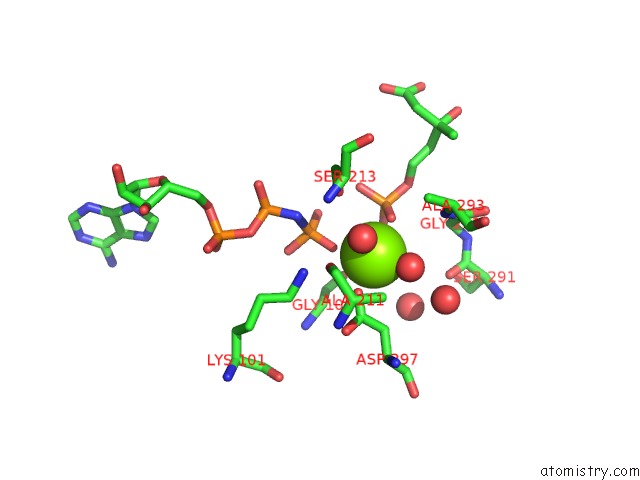
Mono view
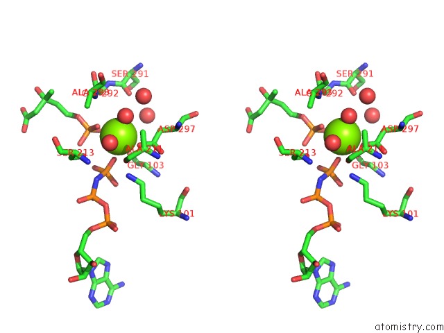
Stereo pair view
Mono view
Stereo pair view
A full contact list of Magnesium with other atoms in the Mg binding
site number 1 of Streptococcus Pneumoniae Phosphomevalonate Kinase in Complex with Phosphomevalonate and Amppnp within 5.0Å range:
|
Reference:
J.L.Andreassi,
M.W.Vetting,
P.W.Bilder,
S.L.Roderick,
T.S.Leyh.
Structure of the Ternary Complex of Phosphomevalonate Kinase: the Enzyme and Its Family Biochemistry V. 48 6461 2009.
ISSN: ISSN 0006-2960
PubMed: 19485344
DOI: 10.1021/BI900537U
Page generated: Wed Aug 14 14:53:44 2024
ISSN: ISSN 0006-2960
PubMed: 19485344
DOI: 10.1021/BI900537U
Last articles
Zn in 9J0NZn in 9J0O
Zn in 9J0P
Zn in 9FJX
Zn in 9EKB
Zn in 9C0F
Zn in 9CAH
Zn in 9CH0
Zn in 9CH3
Zn in 9CH1