Magnesium »
PDB 3gnz-3gx2 »
3gv8 »
Magnesium in PDB 3gv8: Human Dna Polymerase Iota in Complex with T Template Dna and Incoming Dgtp
Enzymatic activity of Human Dna Polymerase Iota in Complex with T Template Dna and Incoming Dgtp
All present enzymatic activity of Human Dna Polymerase Iota in Complex with T Template Dna and Incoming Dgtp:
2.7.7.7;
2.7.7.7;
Protein crystallography data
The structure of Human Dna Polymerase Iota in Complex with T Template Dna and Incoming Dgtp, PDB code: 3gv8
was solved by
K.N.Kirouac,
H.Ling,
with X-Ray Crystallography technique. A brief refinement statistics is given in the table below:
| Resolution Low / High (Å) | 23.84 / 2.00 |
| Space group | P 65 2 2 |
| Cell size a, b, c (Å), α, β, γ (°) | 98.070, 98.070, 203.380, 90.00, 90.00, 120.00 |
| R / Rfree (%) | 21.8 / 24.8 |
Magnesium Binding Sites:
The binding sites of Magnesium atom in the Human Dna Polymerase Iota in Complex with T Template Dna and Incoming Dgtp
(pdb code 3gv8). This binding sites where shown within
5.0 Angstroms radius around Magnesium atom.
In total 2 binding sites of Magnesium where determined in the Human Dna Polymerase Iota in Complex with T Template Dna and Incoming Dgtp, PDB code: 3gv8:
Jump to Magnesium binding site number: 1; 2;
In total 2 binding sites of Magnesium where determined in the Human Dna Polymerase Iota in Complex with T Template Dna and Incoming Dgtp, PDB code: 3gv8:
Jump to Magnesium binding site number: 1; 2;
Magnesium binding site 1 out of 2 in 3gv8
Go back to
Magnesium binding site 1 out
of 2 in the Human Dna Polymerase Iota in Complex with T Template Dna and Incoming Dgtp
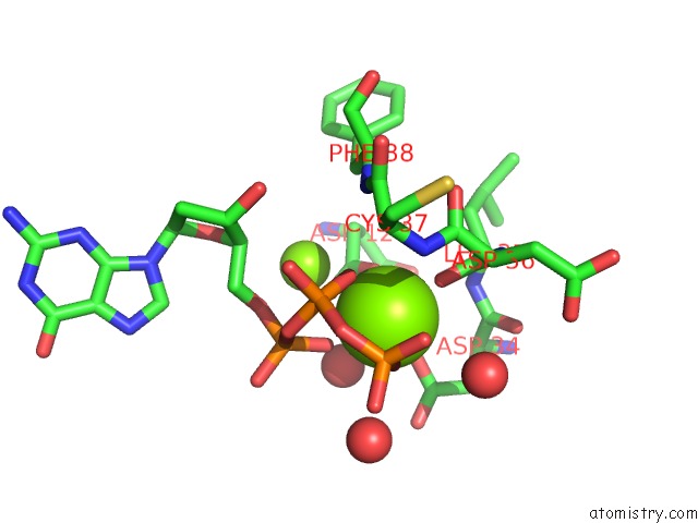
Mono view
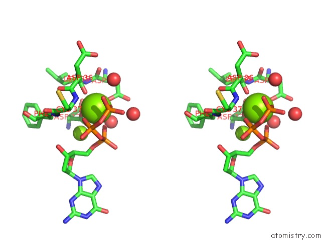
Stereo pair view
Mono view
Stereo pair view
A full contact list of Magnesium with other atoms in the Mg binding
site number 1 of Human Dna Polymerase Iota in Complex with T Template Dna and Incoming Dgtp within 5.0Å range:
|
Magnesium binding site 2 out of 2 in 3gv8
Go back to
Magnesium binding site 2 out
of 2 in the Human Dna Polymerase Iota in Complex with T Template Dna and Incoming Dgtp
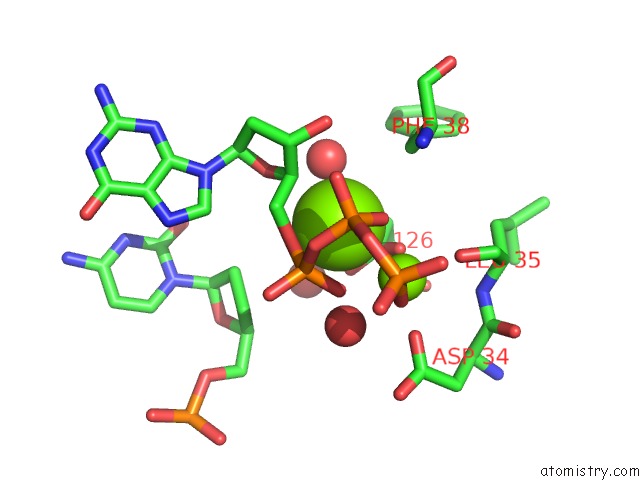
Mono view
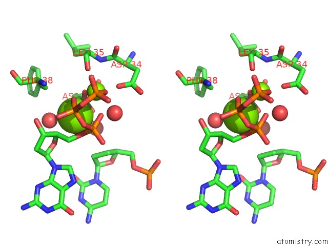
Stereo pair view
Mono view
Stereo pair view
A full contact list of Magnesium with other atoms in the Mg binding
site number 2 of Human Dna Polymerase Iota in Complex with T Template Dna and Incoming Dgtp within 5.0Å range:
|
Reference:
K.N.Kirouac,
H.Ling.
Structural Basis of Error-Prone Replication and Stalling at A Thymine Base By Human Dna Polymerase Iota Embo J. V. 28 1644 2009.
ISSN: ISSN 0261-4189
PubMed: 19440206
DOI: 10.1038/EMBOJ.2009.122
Page generated: Wed Aug 14 14:57:35 2024
ISSN: ISSN 0261-4189
PubMed: 19440206
DOI: 10.1038/EMBOJ.2009.122
Last articles
Ca in 5VERCa in 5VEQ
Ca in 5VE9
Ca in 5VEP
Ca in 5V9U
Ca in 5VCO
Ca in 5VCN
Ca in 5VC1
Ca in 5VA9
Ca in 5VBJ