Magnesium »
PDB 3gnz-3gx2 »
3gwg »
Magnesium in PDB 3gwg: Crystal Structure of Chey of Helicobacter Pylori
Protein crystallography data
The structure of Crystal Structure of Chey of Helicobacter Pylori, PDB code: 3gwg
was solved by
K.H.Lam,
T.K.Ling,
S.W.Au,
with X-Ray Crystallography technique. A brief refinement statistics is given in the table below:
| Resolution Low / High (Å) | 19.37 / 1.80 |
| Space group | C 1 2 1 |
| Cell size a, b, c (Å), α, β, γ (°) | 70.320, 38.476, 38.856, 90.00, 106.95, 90.00 |
| R / Rfree (%) | 14.7 / 18.6 |
Magnesium Binding Sites:
The binding sites of Magnesium atom in the Crystal Structure of Chey of Helicobacter Pylori
(pdb code 3gwg). This binding sites where shown within
5.0 Angstroms radius around Magnesium atom.
In total 2 binding sites of Magnesium where determined in the Crystal Structure of Chey of Helicobacter Pylori, PDB code: 3gwg:
Jump to Magnesium binding site number: 1; 2;
In total 2 binding sites of Magnesium where determined in the Crystal Structure of Chey of Helicobacter Pylori, PDB code: 3gwg:
Jump to Magnesium binding site number: 1; 2;
Magnesium binding site 1 out of 2 in 3gwg
Go back to
Magnesium binding site 1 out
of 2 in the Crystal Structure of Chey of Helicobacter Pylori
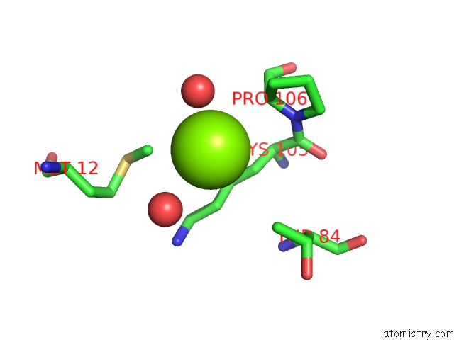
Mono view
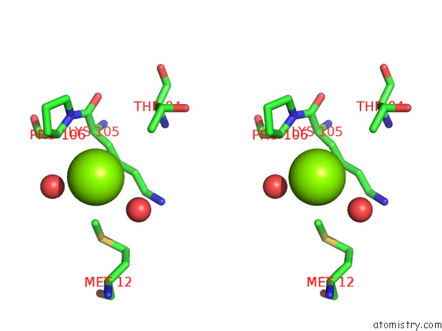
Stereo pair view
Mono view
Stereo pair view
A full contact list of Magnesium with other atoms in the Mg binding
site number 1 of Crystal Structure of Chey of Helicobacter Pylori within 5.0Å range:
|
Magnesium binding site 2 out of 2 in 3gwg
Go back to
Magnesium binding site 2 out
of 2 in the Crystal Structure of Chey of Helicobacter Pylori
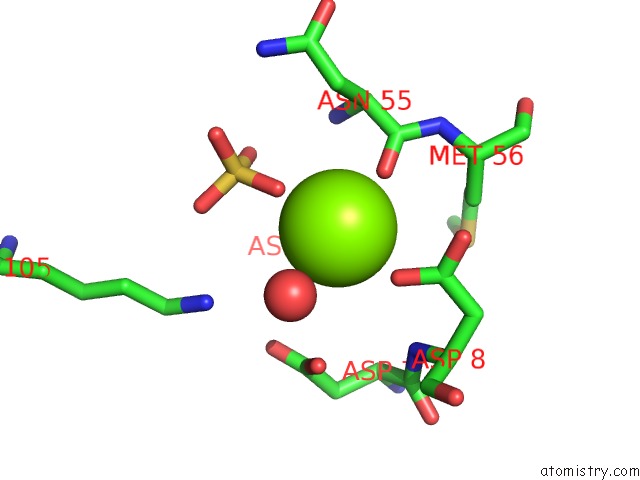
Mono view
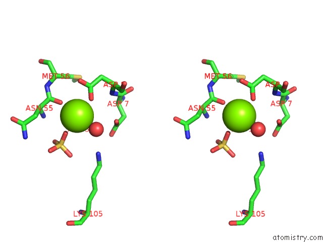
Stereo pair view
Mono view
Stereo pair view
A full contact list of Magnesium with other atoms in the Mg binding
site number 2 of Crystal Structure of Chey of Helicobacter Pylori within 5.0Å range:
|
Reference:
K.H.Lam,
T.K.Ling,
S.W.Au.
Crystal Structure of Activated CHEY1 From Helicobacter Pylori. J.Bacteriol. V. 192 2324 2010.
ISSN: ISSN 0021-9193
PubMed: 20207758
DOI: 10.1128/JB.00603-09
Page generated: Wed Aug 14 14:58:17 2024
ISSN: ISSN 0021-9193
PubMed: 20207758
DOI: 10.1128/JB.00603-09
Last articles
Ca in 5VR0Ca in 5VPL
Ca in 5VPH
Ca in 5VMS
Ca in 5VPG
Ca in 5VOF
Ca in 5VMA
Ca in 5VOE
Ca in 5VLL
Ca in 5VMQ